. 2004 Feb 2;199(3):381-98.
doi: 10.1084/jem.20030132.

Pathogenic profiles and molecular signatures of antinuclear autoantibodies rescued from NZM2410 lupus mice

Affiliations

Pathogenic profiles and molecular signatures of antinuclear autoantibodies rescued from NZM2410 lupus mice

Zhiyan Liang et al. J Exp Med. .

Abstract

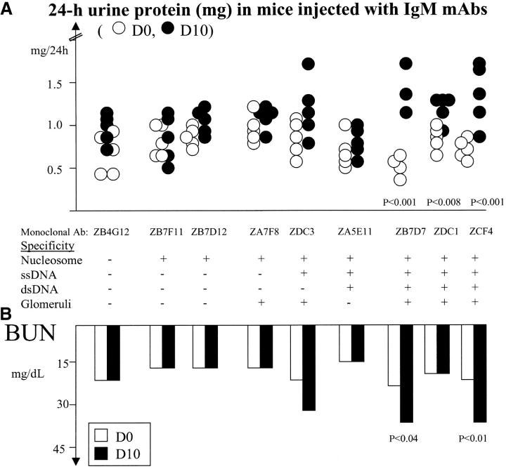
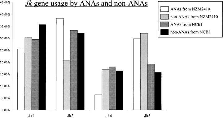
Two outstanding questions concerning antinuclear antibodies (ANAs) in lupus involve their pathogenic potential and their molecular signatures. To address these questions, a panel of 56 antinuclear and 47 nonnuclear binding monoclonal antibodies was rescued from four seropositive NZM2410 lupus mice. The monoclonals varied in their reactivity to nucleosomes, ssDNA, dsDNA, and glomerular substrate. A large fraction of the antibodies demonstrated apparent polyreactivity (to DNA, histones, and glomerular antigens) due to bound, DNase-1 sensitive nuclear antigenic bridges. Although nephrophilic immunoglobulin (Ig) M and IgG antibodies were the most pathogenic, the dsDNA-binding antibodies were modestly so; in contrast, antinucleosome antibodies were clearly not pathogenic. Compared with the nonnuclear antigen-binding monoclonal antibodies rescued from the same mice, ANAs exhibited increased utilization of VH5/7183 genes and highly cationic heavy chain (HC) CDR3 regions. Most intriguingly, the CDR3 regions of the ANAs exhibited alternating arginine/lysine peaks at H96, H98, and H100, with neutral troughs at H95, H97, and H99. To summarize, glomerular-binding anti-dsDNA antibodies appear to be the most pathogenic variety of lupus autoantibodies. The presence of an alternating charge pattern in their HC CDR3 regions appears to be a prominent hallmark of ANAs.


Figures

Figure 1.
Figure 1.
Nuclear antigenic bridges facilitate histone binding by anti-DNA Abs. Six representative anti-dsDNA/antihistone dual-binding Abs were subjected to DNase-I or sham treatment as detailed in Materials and Methods. Likewise, the histone substrate (i.e., “Antigen”) was also subjected to DNase-I or sham treatment. All Abs were tested for histone reactivity within the same ELISA plates. Horizontal bars represent the mean histone reactivity within each treatment group. The depicted p-values represent the result of comparing each group with the sham-treated (Ab and Ag) control. All DNase-I–treated Abs retained dsDNA-binding after treatment (not depicted).
Figure 2.
Figure 2.
Glomerular reactivity of NZM2410-derived ANAs. (A) All the NZM2410-derived ANAs listed in Table I were tested for their reactivity to glomerular substrate as detailed in Materials and Methods. The 56 mAbs studied may be categorized into four groups according to their predominant Ag specificity pattern: anti-ssDNA Abs (n = 11), antihistone/nucleosome Abs (n = 14), nonhistone binding anti-dsDNA Abs (n = 10), and Abs that reacted with both histones and DNA (n = 21). In the composite bar graphs, the percentages of Abs within each group that tested positive for nephrophilicity are shown. (+ and ++) The glomerular antigen-specific reactivity (OD) of the respective mAbs (assayed in the linear range of 1–10 μg/ml) was 0.2–0.5, or >0.5-fold higher, respectively, compared with the corresponding OD values recorded for “total Ig,” assayed in parallel. The percentage of dsDNA/histone dual binder Abs that also reacted with glomerular substrate was significantly higher than the corresponding percentages observed among anti-ssDNA ANAs (P < 0.002), antihistone/nucleosome Abs (P < 0.03), and the nonhistone-binding anti-dsDNA Abs (P < 0.013), as determined using the Fisher's exact test. (B) Five representative antiglomerular Abs (that also reacted with dsDNA) were subjected to DNase-I or sham treatment as detailed in Materials and Methods. Likewise, the glomerular substrate (i.e., Antigen) was also subjected to DNase-I or sham treatment. All Abs were tested for glomerular reactivity within the same ELISA plates. Horizontal bars represent the mean glomerular reactivity within each treatment group. The depicted p-values represent the result of comparing each group with the sham-treated (Ab and Ag) control.
Figure 3.
Figure 3.
Renal pathogenicity of NZM2410-derived IgG ANAs. (A) Seven representative NZM2410-derived IgG mAbs were tested for in vivo pathogenicity as detailed in Materials and Methods. Although ZB4D8, ZDB4, and ZB1A7 were IgG1, IgG2b, and IgG3 in isotype, respectively, all other Abs were IgG2a in isotype. The specificity profiles of these Abs that have been tabulated below the figure were obtained from Table I. It should be noted that “+” in this figure simply indicates that the mAb reacts with the respective Ag, independent of the strength of reactivity. Both the beginning (D0, white dots), and ending (D10, black dots) 24-h urine protein levels (measured using metabolic cages) are depicted. Where the D10 proteinuria levels were found to be significantly higher than the corresponding D0 levels (using the Student's t test), the p-values are listed. (B) The 24-h urine protein excretion profiles of the 10 mice injected with IgG anti-dsDNA/glomerular dual binding Abs, ZB1B11 and ZB4D8, are depicted. 24-h urine protein measurements were performed on D0, D3, D7, and D10 after mAb administration. (C) The BUN levels measured on D0 and D10 after administration of the dsDNA/glomerular dual binding Abs, ZB4D8 and ZB1B11, are depicted. Depicted below each column of dots are the p-values calculated when the D0 and D10 BUN values were compared (using the Student's t test), and found to be significantly different. The dotted line refers to the mean D10 BUN level noted in all the other experimental groups of mice, combined.
Figure 4.
Figure 4.
Renal pathogenicity of NZM2410-derived IgM ANAs. (A) Nine representative NZM2410-derived IgM mAbs were tested for in vivo pathogenicity as detailed in Materials and Methods. The specificity profiles of these Abs that have been tabulated below the figure were obtained from Table I. It should be noted that “+” in this figure simply indicates that the mAb does react with the respective Ag, independent of the strength of reactivity. Both the beginning (D0, white dots) and ending (D10, black dots) 24-h urine protein levels (measured using metabolic cages) are depicted. Where the D10 proteinuria levels were found to be significantly higher than the corresponding D0 levels (using the Student's t test), the p-values are listed. (B) Depicted below are the BUN levels measured on D0 and D10 after Ab administration. Depicted below each column of dots are the p-values (calculated using the Student's t test), where the D0 and D10 BUN values were compared and found to be significantly different.
Figure 5.
Figure 5.
Jk usage frequencies among ANAs and non-ANAs. Indicated are the LC Jk usage frequencies of NZM2410-derived ANAs (n = 46), NZM2410-derived non-ANAs (n = 40), previously documented ANAs (from NCBI/GenBank/EMBL/DDBJ; n = 264), and non-ANAs (from NCBI/GenBank/EMBL/DDBJ; n = 145). The NCBI collection of ANAs and non-ANAs represents two new databases of LC sequences recently constructed and analyzed. The control ANAs represent 264 previously documented ANA LC sequences, drawn from 35 primary works, from which clonal replicates have been removed; they consisted of 139 anti-ssDNA, 103 anti-dsDNA, and 22 antinucleosome Abs (26). The NCBI/GenBank/EMBL/DDBJ “non-ANAs” represent the LC sequences of 145 non-ANAs (with known antigen specificities) drawn from the NCBI/GenBank/EMBL/DDBJ database, with no overlapping target antigen specificities. Importantly, all clonal replicates have been removed from all four of the databases studied, so as to minimize any potential bias due to multi-member clones. The frequencies of Jk5 among the NZM2410-derived ANAs and non-ANAs were significantly higher (P < 0.013 and P < 0.009, respectively) when compared with the corresponding frequencies observed among the NCBI-derived ANAs and non-ANAs.
Figure 6.
Figure 6.
pI profiles across HC CDR3 regions of NZM2410-derived ANAs and non-ANAs. The mean pI value (isoelectric point) at each HC CDR3 position (from H95 to H100a) was calculated as detailed in Materials and Methods. As evident from Table III, few Abs had CDR3 residues beyond position H100a. The mean pI values observed among the NZM2410-derived ANAs (n = 49) and non-ANAs (n = 40) are plotted. Importantly, all clonal replicates have been removed from both databases. The pI values of the ANAs were systematically compared with the pI values of the non-ANAs at each indicated CDR position using the Student's t test (*, P < 0.05; **, P < 0.01).
Figure 7.
Figure 7.
The distribution of “R/K” and “D/E” residues in the HC CDR3 regions of ANAs and non-ANAs. In A, the respective frequencies of R/K and D/E residues at each of the HC CDR3 positions, H95– H100a, among the NZM2410-derived ANAs (n = 49) and non-ANAs (n = 40) are depicted. Differences (between ANAs and non-ANAs) that attained statistical significance are denoted (*, P < 0.05; **, P < 0.01). The solid line arrowhead indicates the alternating frequency peaks of cationicity between H96 and H98 of ANAs; the dotted line arrowhead indicates the alternating peaks of anionicity between H96 and H98 among the non-ANAs. There were too few sequences with CDR3 positions extending beyond H100a (Table III). In B, a similar analysis was performed using previously reported ANAs (n = 269) and non-ANAs (n = 3,600) (for review see reference 25). These control ANAs represent 269 previously documented ANAs drawn from 35 primary works, from which clonal replicates have been removed; they consisted of 143 anti-ssDNA, 103 anti-dsDNA, and 23 antinucleosome Abs (25). The control “non-ANAs” represent the HCs of all Abs deposited in the Kabat database. Differences that attained statistical significance are denoted (*, P < 0.05; **, P < 0.01; ***, P < 0.001).
Figure 8.
Figure 8.
Somatic variations between members of antibody clones. 11 NZM2410-derived monoclonal ANAs are depicted that belonged to five independent clones, A–E, as indicated in Tables I, III, and IV. These clones have been labeled the same way as in Table I, except that the mouse origin identifiers (e.g., “ZA”, “ZB,” etc.) have been omitted. DsDNA-reactive clones are shaded in gray, whereas exclusive nucleosome binders are left unshaded. (top) The HC somatic mutations relative to the closest HC germline gene (indicated in oval labels with dotted borders) are depicted. (bottom) The LC somatic mutations relative to the closest LC germline gene (indicated in oval-shaped labels with dotted borders) are depicted. Thus, in clone “B,” mAbs 7H10 and 3F7 vary from the J558 germline gene, V23, by 6 or 10 residues, respectively, whereas they both bear the same unmutated LC Vk1 germline gene, bb1. Interestingly, whereas the first-listed two members of Clone D, 7F11 and 2D7, possessed a mutated RF Vk germline gene, the third member possessed an entirely different LC gene that differed from the Vk9 germline gene, ba9, by nine somatic mutations (not depicted).

Similar articles

Cited by

References

    1. Kotzin, B.L. 1996. Systemic lupus erythematosus. Cell. 85:303–306. - PubMed
    1. Hahn, B.H. 1998. Antibodies to DNA. N. Engl. J. Med. 338:1359–1368. - PubMed
    1. Pisetsky, D.H. 2000. Anti-DNA and autoantibodies. Curr. Opin. Rheumatol. 12:364–368. - PubMed
    1. Lefkowith, J.B., and G.S. Gilkeson. 1996. Nephritogenic autoantibodies in lupus. Current concepts and continuing controversies. Arthritis Rheum. 39:894–903. - PubMed
    1. Vlahakos, D.V., M.H. Foster, S. Adams, M. Katz, A.A. Ucci, K.J. Barrett, S.K. Datta, and M.P. Madaio. 1992. Anti-DNA antibodies form immune deposits at distinct glomerular and vascular sites. Kidney Int. 41:1690–1700. - PubMed

Publication types