. 2004 Feb 2;199(3):399-410.
doi: 10.1084/jem.20032092.

Survivin loss in thymocytes triggers p53-mediated growth arrest and p53-independent cell death

Affiliations

Survivin loss in thymocytes triggers p53-mediated growth arrest and p53-independent cell death

Hitoshi Okada et al. J Exp Med. .

Abstract

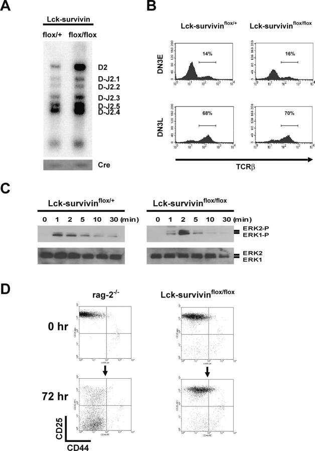
Because survivin-null embryos die at an early embryonic stage, the role of survivin in thymocyte development is unknown. We have investigated the role by deleting the survivin gene only in the T lineage and show here that loss of survivin blocks the transition from CD4- CD8- double negative (DN) thymocytes to CD4+ CD8+ double positive cells. Although the pre-T cell receptor signaling pathway is intact in survivin-deficient thymocytes, the cells cannot respond to its signals. In response to proliferative stimuli, cycling survivin-deficient DN cells exhibit cell cycle arrest, a spindle formation defect, and increased cell death. Strikingly, loss of survivin activates the tumor suppressor p53. However, the developmental defects caused by survivin deficiency cannot be rescued by p53 inactivation or introduction of Bcl-2. These lines of evidence indicate that developing thymocytes depend on the cytoprotective function of survivin and that this function is tightly coupled to cell proliferation but independent of p53 and Bcl-2. Thus, survivin plays a critical role in early thymocyte development.


Figures

Figure 1.
Figure 1.
Tissue-specific targeted disruption of the survivin locus. (A) A portion of the murine wild-type survivin locus (top) showing exons 1–3 (open boxes) and a 10-kb XbaI fragment. The targeting vector was designed to generate floxed exon2 (loxP; triangles), flank the PGK-neo cassette with FRT sequences (ovals), and introduce a new XbaI site (X). The targeted allele contains a diagnostic 7.0-kb XbaI fragment. To generate the conditional allele, the neo cassette was removed by transient expression of Flpe recombinase. Cre-mediated recombination resulted in the deleted allele. The positions of the 5′ flanking probe (A) and neo probe (B) used for genotyping are indicated. (B) Southern blot analyses to identify survivinflox/+ ES cells containing neo (left), ES cells with neo removed (middle), and F2 pups (right). Genomic DNA was digested with XbaI and hybridized with probe A or B. (C) Western blot of survivin protein in Lck-survivinflox/+ and Lck-survivinflox/flox DN thymocytes. Wild-type survivin protein (arrowhead) and a nonspecific band (asterisk) are indicated. Actin, loading control. (D) Detection of deleted survivin allele in DN thymocyte subpopulations. The floxed (flox) and deleted (Δflox) survivin alleles were amplified by PCR using primers a and b shown in A. Genomic DNA samples were prepared from the indicated DN subsets.
Figure 2.
Figure 2.
Flow cytometric and histological analyses of peripheral T cells and thymi of Lck-Cre;survivinflox/flox mice. (A) Survivin mRNA expression in DN cells. Levels of survivin mRNA were analyzed in the indicated DN subsets by RT-PCR. RNA was prepared from DN cells sorted from wild-type thymocytes. (B) Survivin protein expression in DN cells. Surface staining of Lck-survivin+/+ (thick line) and Lck-survivinflox/flox (dotted line) thymocytes with anti-CD25, anti-CD44, and anti-Lin Abs was followed by intracellular staining with anti-survivin Ab. Lin+ cells in the indicated thymocyte subpopulations were electrically gated out. (C) Impaired development of survivin-deficient peripheral T cells and DP thymocytes. Peripheral blood cells from Lck-survivin+/+, Lck-survivinflox/+, and Lck-survivinflox/flox mice were stained with anti-TCRαβ and anti-B220 (top). Thymocytes were stained with anti-CD4 and anti-CD8 (middle), or anti-CD25 and anti-CD44 (bottom), and the Lin population was examined. (D) Reduced size and cellularity of the thymus in Lck-survivinflox/flox mice. Hematoxylin and eosin staining of transverse sections of thymus from Lck-survivinflox/+ (left) and Lck-survivinflox/flox (right) mice. The mutant thymus is much smaller and lacks the typical cortex medulla structure.
Figure 3.
Figure 3.
Normal TCRβ gene rearrangement and pre-TCR signaling in Lck-Cre;survivinflox/flox thymocytes. (A) Normal TCRβ D-J rearrangement. TCRβ Dβ2-Jβ2 recombination in Lck-survivinflox/+ and Lck-survivinflox/flox DN thymocytes was examined by PCR followed by Southern blotting with 32P-labeled oligonucleotide probes as previously described (reference 59). The Cre gene was amplified as an internal control for PCR. (B) Normal TCRβ protein expression. DN3E and DNL3 thymocytes from Lck-survivinflox/+ and Lck-survivinflox/flox mice were surface stained with anti-CD25, anti-CD44, and anti-Lin followed by intracellular staining with TCRβ and flow cytometric analysis. (C) Normal MAPK activation. Lck-survivinflox/+ and Lck-survivinflox/flox DN thymocytes were treated with anti–CD3ε-biotin followed by cross-linking with avidin for the indicated times. Protein lysates were subjected to Western blot analysis using anti–phospho-ERK Abs (top). To control for loading, the blot was stripped and reprobed with anti–ERK Ab (bottom). (D) Impaired in vivo response of survivin-deficient thymocytes to anti-CD3ε. RAG-2−/− (left) and Lck-survivinflox/flox (right) mice were intraperitoneally injected with 100 mg anti-CD3ε. After 72 h, thymocytes were prepared and stained with anti-CD25, anti-CD44, and anti-Lin Abs, and then subjected to flow cytometry. Unlike RAG-2−/− DN3 cells, Lck-survivinflox/flox DN3 cells failed to advance to DN4. Data shown are representative of three independent experiments.
Figure 4.
Figure 4.
Effect of survivin loss on cell death in vivo and in vitro and on thymocyte proliferation. (A) Increased cell death of proliferating survivin-deficient DN cells. DN thymocytes were prepared ex vivo and stained with anti-CD25, anti-CD44, and anti-Lin Ab followed by annexin. The average number of annexin+ cells in each DN subpopulation was assessed by flow cytometry. Results shown are mean ± SD. Annexin+ cells were significantly increased in Lck-survivinflox/flox DN3L and DN4 populations (ANOVA; n = 3). (B) Normal susceptibility of resting survivin-deficient DN cells to various apoptotic stimuli. Lck-survivinflox/flox and RAG-2−/− DN3E cells were treated with etoposide (Etp), dexamethasone (Dex), γ-irradiation (IR), and staurosporine (STS) at the indicated doses. Cell death was evaluated as described in Materials and Methods. Cell viability was normalized to account for spontaneous cell death. Triplicate samples of each treatment in three independent experiments were assayed. Results shown are mean ± SD. (C) Impaired proliferation in vivo in the absence of survivin. Lck-survivinflox/+ and Lck-survivinflox/flox mice were injected with 1 mg BrdU and DN thymocytes were purified and stained with anti-CD25, anti-CD44, anti-BrdU, and 7AAD. The percentages of cells in the G1, S, and G2/M phases were measured by flow cytometry for the indicated DN subsets. Results shown are representative of three independent experiments.
Figure 5.
Figure 5.
Defects in cytokinesis of Lck-survivinflox/flox DN thymocytes. (A) Impaired spindle formation. Purified DN3L and DN4 cells from Lck-survivin+/+ (a) and Lck-survivinflox/flox (b) mice were stained with anti–α-tubulin (green). DNA was visualized with Hoechst 33258 (blue) staining. Data shown are representative of at least three independent preparations per genotype. (B) Severe impairment of spindle assembly in mitotic cells. Spindle assembly in cells from Lck-survivinflox/flox mice was compared at interphase (bottom left) and mitosis (top right). Cells were stained with anti–α-tubulin (green), anti–phospho-histone H3 (p-H3) Ab (red), and Hoechst (blue). a, α-tubulin; b, anti–p-H3 and Hoechst; c, merge. (C) Impaired cytokinesis and localization of Aurora-B kinase. Mitotic DN3L and DN4 cells from Lck-survivin+/+ (a–d) and Lck-survivinflox/flox (e–h) mice were stained with anti–p-H3 Ab (red), anti–Aurora-B kinase Ab (green), and Hoechst (blue). a and e, prometaphase; b and f, metaphase; c and g, anaphase; d and h, telophase/cytokinesis.
Figure 6.
Figure 6.
Induction of p53 and p21 in Lck-survivinflox/flox DN thymocytes. (A) p53 and p21 induction in DN thymocytes. Top: Protein samples prepared from Lck-survivinflox/+ and Lck-survivinflox/flox DN cells were subjected to Western blot analysis using anti-p53 Ab followed by reprobing with anti-actin as a loading control. Bottom: Levels of p21 and HPRT (loading control) mRNA in control and mutant DN cells were determined by RT-PCR. (B) p53 induction in DN subpopulations. Lck-survivinflox/+ and Lck-survivinflox/flox thymocytes were surface stained with anti-CD25, anti-CD44, and anti-Lin followed by intracellular staining with anti-p53 or control IgG. p53 protein in each DN subpopulation was assessed by flow cytometry.
Figure 7.
Figure 7.
Effects of Bcl-2 gain or p53 loss on phenotypes of survivin-deficient thymocytes. (A) Gain of Bcl-2 does not restore DP cells in the absence of survivin. Thymocytes prepared from Lck-survivinflox/+;Bcl-2, Lck-survivinflox/flox, and Lck-survivinflox/flox;Bcl-2 mice were stained with anti-CD4 and anti-CD8 followed by flow cytometric analysis. One experiment representative of four independent trials is shown. (B) Loss of p53 does not rescue DN thymocyte development in the absence of survivin. CD4/CD8 expression was determined as in A for thymocytes from Lck-survivinflox/+;p53−/−, Lck-survivinflox/flox;p53+/−, and Lck-survivinflox/flox;p53−/− mice. (C) Decreased G1 arrest of survivin-deficient DN3L cells in the p53−/− and p21−/− genetic backgrounds. Cell cycle profiles of DN cells prepared from Lck-survivinflox/+, Lck-survivinflox/flox, Lck-survivinflox/flox;p53−/−, and Lck-survivinflox/flox;p21−/− mice were determined as in Fig. 4 B. (D) Increased cell death of survivin-deficient DN cells in the p53−/− and p21 genetic backgrounds. The percentages of annexin+ cells in the indicated DN subpopulations were determined as in Fig. 4 A. Results shown are mean ± SD. Annexin+ cells were significantly increased in p53−/− and p21−/− genetic backgrounds (ANOVA; n = 3).

Similar articles

Cited by

References

    1. Godfrey, D.I., J. Kennedy, T. Suda, and A. Zlotnik. 1993. A developmental pathway involving four phenotypically and functionally distinct subsets of CD3−CD4−CD8− triple-negative adult mouse thymocytes defined by CD44 and CD25 expression. J. Immunol. 150:4244–4252. - PubMed
    1. Dudley, E.C., H.T. Petrie, L.M. Shah, M.J. Owen, and A.C. Hayday. 1994. T cell receptor beta chain gene rearrangement and selection during thymocyte development in adult mice. Immunity. 1:83–93. - PubMed
    1. Levelt, C.N., and K. Eichmann. 1995. Receptors and signals in early thymic selection. Immunity. 3:667–672. - PubMed
    1. von Boehmer, H., I. Aifantis, J. Feinberg, O. Lechner, C. Saint-Ruf, U. Walter, J. Buer, and O. Azogui. 1999. Pleiotropic changes controlled by the pre-T-cell receptor. Curr. Opin. Immunol. 11:135–142. - PubMed
    1. Akashi, K., M. Kondo, U. von Freeden-Jeffry, R. Murray, and I.L. Weissman. 1997. Bcl-2 rescues T lymphopoiesis in interleukin-7 receptor-deficient mice. Cell. 89:1033–1041. - PubMed

Publication types

MeSH terms