. 2004 Feb 2;164(3):385-94.
doi: 10.1083/jcb.200307132.

Krox-20 inhibits Jun-NH2-terminal kinase/c-Jun to control Schwann cell proliferation and death

Affiliations

Krox-20 inhibits Jun-NH2-terminal kinase/c-Jun to control Schwann cell proliferation and death

David B Parkinson et al. J Cell Biol. .

Abstract

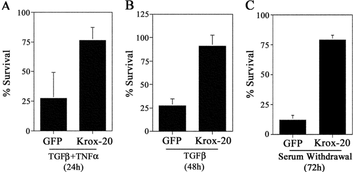
The transcription factor Krox-20 controls Schwann cell myelination. Schwann cells in Krox-20 null mice fail to myelinate, and unlike myelinating Schwann cells, continue to proliferate and are susceptible to death. We find that enforced Krox-20 expression in Schwann cells cell-autonomously inactivates the proliferative response of Schwann cells to the major axonal mitogen beta-neuregulin-1 and the death response to TGFbeta or serum deprivation. Even in 3T3 fibroblasts, Krox-20 not only blocks proliferation and death but also activates the myelin genes periaxin and protein zero, showing properties in common with master regulatory genes in other cell types. Significantly, a major function of Krox-20 is to suppress the c-Jun NH2-terminal protein kinase (JNK)-c-Jun pathway, activation of which is required for both proliferation and death. Thus, Krox-20 can coordinately control suppression of mitogenic and death responses. Krox-20 also up-regulates the scaffold protein JNK-interacting protein 1 (JIP-1). We propose this as a possible component of the mechanism by which Krox-20 regulates JNK activity during Schwann cell development.


Figures

Figure 1.
Figure 1.
Krox-20 inhibits DNA synthesis in Schwann cells. (A) The proliferation of Schwann cells, as measured by BrdU incorporation, in response to NRG-1 is inhibited by Krox-20. Schwann cells retrovirally infected with empty vector (BP) or vector expressing Krox-20 (K20) were treated with medium alone (Control) or with medium supplemented with 20 ng/ml NRG-1. The error bars represent one SD of the mean. (B) Inhibition of BrdU incorporation in cells expressing Krox-20. Cells infected with adenovirus coexpressing GFP and Krox-20 in control (−NRG-1) and 20 ng/ml NRG-1–treated cells. Note that GFP/Krox-20–positive cells are BrdU negative. Conversely, BrdU-positive cells are GFP negative (arrows). Bar, 20 μm. (C and D) Krox-20 expression inhibits cyclin D1 induction in response to NRG-1. (C) The graph shows percentage of adenovirally infected GFP/cyclin D1–positive cells 6 h after addition of 20 ng/ml NRG-1 in GFP control (GFP) and GFP/Krox-20 (Krox-20)–expressing cells. The error bars represent one SD of the mean. (D) Western blot of cyclin D1 in GFP control (GFP) and GFP/Krox-20 (Krox-20) infected Schwann cells exposed to DM alone (Con) or DM containing 20 ng/ml NRG-1 for 6 h. (E–I) Krox-20 expression increases levels of p27 protein in Schwann cells. (E–H) Immunolabeling of GFP (E and G) and GFP/Krox-20 (F and H) infected Schwann cells with p27 antibody. Arrows indicate GFP/Krox-20 cells with elevated p27 protein. Bar, 20 μm. (I) Western blot using p27 antibody of Schwann cells infected with either empty vector (BP) or Krox-20 (K20)–expressing retrovirus.
Figure 2.
Figure 2.
Krox-20 inhibits apoptosis of Schwann cells. (A and B) Krox-20 inhibits TGFβ-dependent apoptosis of Schwann cells. Survival assays for immunopanned Schwann cells infected with GFP or GFP/Krox-20 (Krox-20)–expressing adenovirus. Cells were treated for 24 h with TGFβ + TNFα (A) or TGFβ alone (B) for 48 h and the number of surviving GFP-positive cells counted. (C) Krox-20 inhibits Schwann cell death after serum withdrawal. Survival assay of cells infected with GFP or GFP/Krox-20 (Krox-20)–expressing adenovirus. Surviving GFP-positive cells were counted 72 h after serum withdrawal. The error bars represent one SD of the mean.
Figure 3.
Figure 3.
Krox-20 regulates proliferation, survival and myelin gene expression in 3T3 fibroblasts. (A) Krox-20 expression inhibits PDGF-induced proliferation of Swiss 3T3 cells. Shown are the percentage of BrdU-positive cells in empty vector (BP) and Krox-20–expressing (K20) cells in the absence (Control) and presence (+PDGF) of 5 ng/ml PDGF. The error bars represent one SD of the mean. (B) Krox-20 expression in Swiss 3T3 cells inhibits cyclin D1 induction in response to PDGF. Shown are control (BP) and Krox-20 (K20) infected cells, treated for 6 h with 5 ng/ml PDGF. The cells were double immunolabeled with Krox-20 (K20) and cyclin D1 (CycD1) antibodies, and counterstained with Hoechst dye (Ho). Bar, 20 μm. (C) Krox-20 expression inhibits apoptosis of NIH 3T3 cells in serum-free medium. The graph shows the percentage of TUNEL-positive cells in control (BP) and Krox-20 (K20)–expressing cells 48 h after serum withdrawal. The error bars represent one SD of the mean. (D–G) Krox-20 activates periaxin and P0 myelin protein expression in Swiss 3T3 cells. Swiss 3T3 cells were retrovirally infected with either empty vector (BP; D and F) or Krox-20 (K20; E and G), and immunolabeled with antibody against periaxin (Prx; D and E) or P0 (F and G). Cells were counterstained with Hoechst dye (Ho) to reveal nuclei. Bar, 20 μm. (H) Western blot of control (BP) or Krox-20 (K20) infected cells showing elevated periaxin and P0 protein levels.
Figure 3.
Figure 3.
Krox-20 regulates proliferation, survival and myelin gene expression in 3T3 fibroblasts. (A) Krox-20 expression inhibits PDGF-induced proliferation of Swiss 3T3 cells. Shown are the percentage of BrdU-positive cells in empty vector (BP) and Krox-20–expressing (K20) cells in the absence (Control) and presence (+PDGF) of 5 ng/ml PDGF. The error bars represent one SD of the mean. (B) Krox-20 expression in Swiss 3T3 cells inhibits cyclin D1 induction in response to PDGF. Shown are control (BP) and Krox-20 (K20) infected cells, treated for 6 h with 5 ng/ml PDGF. The cells were double immunolabeled with Krox-20 (K20) and cyclin D1 (CycD1) antibodies, and counterstained with Hoechst dye (Ho). Bar, 20 μm. (C) Krox-20 expression inhibits apoptosis of NIH 3T3 cells in serum-free medium. The graph shows the percentage of TUNEL-positive cells in control (BP) and Krox-20 (K20)–expressing cells 48 h after serum withdrawal. The error bars represent one SD of the mean. (D–G) Krox-20 activates periaxin and P0 myelin protein expression in Swiss 3T3 cells. Swiss 3T3 cells were retrovirally infected with either empty vector (BP; D and F) or Krox-20 (K20; E and G), and immunolabeled with antibody against periaxin (Prx; D and E) or P0 (F and G). Cells were counterstained with Hoechst dye (Ho) to reveal nuclei. Bar, 20 μm. (H) Western blot of control (BP) or Krox-20 (K20) infected cells showing elevated periaxin and P0 protein levels.
Figure 4.
Figure 4.
Krox-20 does not inactivate NRG-1 or TGFβ signaling. (A) Western blot analysis of control (GFP) and Krox-20 adenovirally infected Schwann cells, showing levels of ErbB2 and ErbB3 protein, together with activation (i.e., phosphorylation) of ERK1/2 and Akt in DM only and 10 min after addition of 20 ng/ml NRG-1. Shown also are the corresponding levels of periaxin in control cells and in cells expressing Krox-20. (B) NRG-1 receptor levels are not altered in Krox-20−/− nerves. Western blot of ErbB2 and ErbB3 in sciatic nerve from P10 wild-type (+/+), Krox-20 heterozygous (+/−) and Krox-20 null (−/−) animals. (C–F) SMAD2 and SMAD4 proteins translocate normally to the nucleus in response to TGFβ in Krox-20–expressing Schwann cells. Note intense nuclear labeling in cells infected with Krox-20–expressing adenovirus, treated for 1 h with 10 ng/ml TGFβ1 and immunolabeled with antibodies to either SMAD2 (C and D) or SMAD4 (E and F). Bar, 20 μm.
Figure 5.
Figure 5.
Krox-20 inhibits the JNK–c-Jun pathway in vitro and in vivo. (A) Western blot (long exposure) showing suppression of phospho–c-Jun (P-cJun), c-Jun protein (cJun), and phospho-JNK1/2 (P-JNK1/2) in cells infected with Krox-20/GFP adenovirus for 48 h and maintained in DM alone. GFP indicates control cells infected with empty vector. (B–E) Krox-20 expression in Schwann cells inhibits NRG-1– (B and C) and TGFβ-dependent (D and E) activation of the JNK–c-Jun pathway. (B and D) Western blot of Schwann cells, 20 h after adenoviral infection, treated with 20 ng/ml NRG-1 (B) or 10 ng/ml TGFβ (D). Note inhibition of c-Jun phosphorylation by Krox-20 after growth factor addition compared with GFP control, whereas c-Jun protein levels are unaffected. (C and E) Western of Schwann cells 48 h after infection, treated with NRG-1 (C) or TGFβ (E). At this time point, both phospho–c-Jun and c-Jun proteins are suppressed by Krox-20 as compared with control cells. Either β-tubulin or GAPDH were used as loading controls. (F–M) c-Jun and Ser63 phospho–c-Jun immunolabeling of Schwann cells infected with either GFP (F, H, J, and L) or GFP/Krox-20 (G, I, K, and M) and treated 48 h after infection with NRG-1 for 30 min. Both c-Jun and phospho–c-Jun levels are reduced in response to NRG-1 in Krox-20–expressing cells compared with GFP control infected cells (arrows). (N and O) Teased preparation of sciatic nerve at E17, before myelination double labeled with c-Jun and Krox-20 antibodies. Hoechst nuclear dye (Ho) is used to visualize nuclei. Note that most/all nuclei contain c-Jun, whereas Krox-20 is not yet present. (P and Q) Teased preparation of sciatic nerve at P1 when many cells are myelinating. The nerves are double immunolabeled with Krox-20 (K20) and c-Jun (P) or phospho–c-Jun (PSer63c-Jun; Q) antibodies. Note that nuclei expressing Krox-20 do not contain c-Jun or phospho–c-Jun. Bar, 20 μm.
Figure 6.
Figure 6.
Inhibition of the JNK pathway inhibits NRG-1–dependent proliferation of Schwann cells. (A) Western blot showing inhibition of NRG-1–induced JNK and c-Jun phosphorylation by 30 μM SP600125 (SP6). (B) Percentage of BrdU-positive Schwann cells in DM only (Cont.) and in medium containing 20 ng/ml NRG-1 in the absence or presence of increasing concentrations of the JNK inhibitor SP600125 as indicated. The error bars represent one SD of the mean. (C) Expression of the JBD of JIP-1 inhibits NRG-1–induced c-Jun phosphorylation, but not ERK1/2 activation. (D) Percentage of BrdU-positive Schwann cells infected with control virus expressing GFP only (GFP) or JBD-expressing adenovirus in the absence (Cont.) or presence of 20 ng/ml NRG-1. The error bars represent one SD of the mean.
Figure 7.
Figure 7.
Expression of MEKK1 or MKK7D and the resulting activation of c-Jun phosphorylation restores vulnerability to death in cells expressing Krox-20. (A) Western blot showing induction of c-Jun phosphorylation in Schwann cells coexpressing Krox-20 and either MEKK1 (K20/MEKK1) or MKK7D (K20/MKK7D) as compared with control (K20/LacZ) cells. (B) Serum withdrawal survival assay of Krox-20/LacZ, Krox-20/MEKK1 or Krox-20/MKK7D–expressing Schwann cells. The death protection offered by Krox-20 alone (Fig. 2 C) is not seen in Krox-20/MEKK1 or Krox-20/MKK7D– expressing cells. The error bars represent one SD of the mean.
Figure 8.
Figure 8.
Regulation of JIP-1 during sciatic development. (A) Western blot of GFP and Krox-20/GFP (K20) adenovirally infected Schwann cells showing increased levels of JIP-1 in Krox-20–expressing cells. (B) JIP-1 protein levels are reduced, and c-Jun levels increased, in sciatic nerve from P9 and P12 Krox-20 null animals (−/−) compared with wild type (+/+). (C) Semi-quantitative PCR analysis of JIP-1 mRNA expression in sciatic nerve during development from embryonic day 14 (E14) to P12 and at P12, 5 d after nerve cut (P12/CUT). (D) Expression of JIP-1 inhibits c-Jun phosphorylation in Schwann cells. Shown are the percentage of Ser63 phospho–c-Jun–positive cells for cells cotransfected with either GFP-expressing plasmid, for identification of transfected cells, plus empty vector (GFP/EV) or GFP plus JIP-1 (GFP/JIP-1)–expressing plasmids. The error bars represent one SD of the mean. (E) Western blot showing that, unlike Krox-20, JIP-1 expression does not reduce c-Jun protein levels in Schwann cells. (F and G) Immunolabeling of P12 sciatic nerve showing phase (F) and JIP-1 immunofluorescence (G). Arrows indicate localization of JIP-1 in the paranodal loops of myelinating Schwann cells.
Figure 8.
Figure 8.
Regulation of JIP-1 during sciatic development. (A) Western blot of GFP and Krox-20/GFP (K20) adenovirally infected Schwann cells showing increased levels of JIP-1 in Krox-20–expressing cells. (B) JIP-1 protein levels are reduced, and c-Jun levels increased, in sciatic nerve from P9 and P12 Krox-20 null animals (−/−) compared with wild type (+/+). (C) Semi-quantitative PCR analysis of JIP-1 mRNA expression in sciatic nerve during development from embryonic day 14 (E14) to P12 and at P12, 5 d after nerve cut (P12/CUT). (D) Expression of JIP-1 inhibits c-Jun phosphorylation in Schwann cells. Shown are the percentage of Ser63 phospho–c-Jun–positive cells for cells cotransfected with either GFP-expressing plasmid, for identification of transfected cells, plus empty vector (GFP/EV) or GFP plus JIP-1 (GFP/JIP-1)–expressing plasmids. The error bars represent one SD of the mean. (E) Western blot showing that, unlike Krox-20, JIP-1 expression does not reduce c-Jun protein levels in Schwann cells. (F and G) Immunolabeling of P12 sciatic nerve showing phase (F) and JIP-1 immunofluorescence (G). Arrows indicate localization of JIP-1 in the paranodal loops of myelinating Schwann cells.

Similar articles

Cited by

References

    1. Awatramani, R., S. Shumas, J. Kamholz, and S.S. Scherer. 2002. TGFbeta1 modulates the phenotype Schwann cells at the transcriptional level. Mol. Cell. Neurosci. 19:307–319. - PubMed
    1. Archelos, J.J., K. Roggenbuck, J. Schneider-Schaulies, C. Linington, K.V. Toyka, and H.P. Hartung. 1993. Production and characterization of monoclonal antibodies to the extracellular domain of P0. J. Neurosci. Res. 35:46–53. - PubMed
    1. Bennett, B.L., D.T. Sasaki, B.W. Murray, E.C. O'Leary, S.T. Sakata, W. Xu, J.C. Leisten, A. Motiwala, S. Pierce, Y. Satoh, et al. 2001. SP600125, an anthrapyrazolone inhibitor of Jun N-terminal kinase. Proc. Natl. Acad. Sci. USA. 98:13681–13686. - PMC - PubMed
    1. Bonny, C., A. Oberson, M. Steinmann, D.F. Schorderet, P. Nicod, and G. Waeber. 2000. IB1 reduces cytokine-induced apoptosis of insulin-secreting cells. J. Biol. Chem. 275:16466–16472. - PubMed
    1. Brockes, J.P., K.L. Fields, and M.C. Raff. 1979. Studies on cultured rat Schwann cells. I. Establishment of purified populations from cultures of peripheral nerve. Brain Res. 165:105–118. - PubMed

Publication types

MeSH terms

Substances