. 2004 Feb 2;164(3):395-405.
doi: 10.1083/jcb.200309159.

A novel LIM protein Cal promotes cardiac differentiation by association with CSX/NKX2-5

Affiliations

A novel LIM protein Cal promotes cardiac differentiation by association with CSX/NKX2-5

Hiroshi Akazawa et al. J Cell Biol. .

Abstract

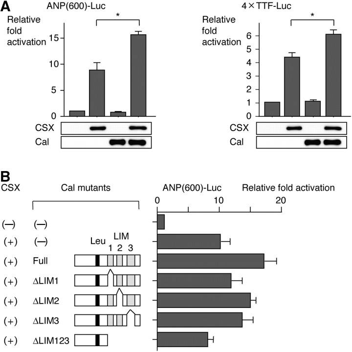
The cardiac homeobox transcription factor CSX/NKX2-5 plays an important role in vertebrate heart development. Using a yeast two-hybrid screening, we identified a novel LIM domain-containing protein, named CSX-associated LIM protein (Cal), that interacts with CSX/NKX2-5. CSX/NKX2-5 and Cal associate with each other both in vivo and in vitro, and the LIM domains of Cal and the homeodomain of CSX/NKX2-5 were necessary for mutual binding. Cal itself possessed the transcription-promoting activity, and cotransfection of Cal enhanced CSX/NKX2-5-induced activation of atrial natriuretic peptide gene promoter. Cal contained a functional nuclear export signal and shuttled from the cytoplasm into the nucleus in response to calcium. Accumulation of Cal in the nucleus of P19CL6 cells promoted myocardial cell differentiation accompanied by increased expression levels of the target genes of CSX/NKX2-5. These results suggest that a novel LIM protein Cal induces cardiomyocyte differentiation through its dynamic intracellular shuttling and association with CSX/NKX2-5.


Figures

Figure 1.
Figure 1.
Deduced amino acid sequence and expression of Cal in embryonic and adult mouse tissues. (A) Deduced amino acid sequence of mouse Cal (GenBank/EMBL/DDBJ accession no. AF513359). LIM domains of Cal are indicated by open boxes, and the leucine-rich sequence is indicated by a gray box. Stretches of consecutive proline residues are underlined. (B) Northern blot analysis of Cal expression in adult mouse tissues. (C) Section in situ hybridization analysis of Cal expression during embtyogenesis. Coronal sections at the level of the heart at E10.5 and E13.5 show Cal expression in a variety of tissues including ventricles, atria, cardiac cushion, and aorta. Ao, aorta; AVC, atrioventricular canal; CC, cardiac cushion; DAo, dorsal aorta; Fo, fourth ventricle; In, intestine; LA, left atrium; Li, liver; Lu, lung; LV, left ventricle; RA, right atrium; RV, right ventricle; St, stomach; Te, telencephalon; Th, third ventricle; V, ventricles; Ve, vertebral column.
Figure 1.
Figure 1.
Deduced amino acid sequence and expression of Cal in embryonic and adult mouse tissues. (A) Deduced amino acid sequence of mouse Cal (GenBank/EMBL/DDBJ accession no. AF513359). LIM domains of Cal are indicated by open boxes, and the leucine-rich sequence is indicated by a gray box. Stretches of consecutive proline residues are underlined. (B) Northern blot analysis of Cal expression in adult mouse tissues. (C) Section in situ hybridization analysis of Cal expression during embtyogenesis. Coronal sections at the level of the heart at E10.5 and E13.5 show Cal expression in a variety of tissues including ventricles, atria, cardiac cushion, and aorta. Ao, aorta; AVC, atrioventricular canal; CC, cardiac cushion; DAo, dorsal aorta; Fo, fourth ventricle; In, intestine; LA, left atrium; Li, liver; Lu, lung; LV, left ventricle; RA, right atrium; RV, right ventricle; St, stomach; Te, telencephalon; Th, third ventricle; V, ventricles; Ve, vertebral column.
Figure 2.
Figure 2.
Complex formation between CSX/NKX2-5 and Cal. (A) Coimmunoprecipitation of CSX/NKX2-5 and Cal in transfected COS7 cells. Immunoprecipitates with anti-FLAG antibody were separated by SDS-PAGE and immunoblotted with anti-HA antibody (top). The same blot was reprobed with anti-FLAG antibody to confirm the presence of FLAG-tagged Cal (bottom). (B) GST pull-down assay for mapping of a region in CSX/NKX2-5 required for binding to Cal. In vitro–translated CSX/NKX2-5 and its mutants labeled with 35S were incubated with GST-Cal immobilized on glutathione-Sepharose beads, and bound proteins were separated by SDS-PAGE and visualized by autoradiography. The arrow indicates the CSX/NKX2-5 protein bound to GST-Cal. A CSX/NKX2-5 mutant lacking the homeodomain did not associate with Cal (arrowhead), whereas a CSX/NKX2-5 mutant containing only the homeodomain did associate. HD, homeodomain. (C) GST pull-down assay for mapping of a region in Cal required for binding to CSX/NKX2-5. In vitro–translated Cal and its mutants labeled with 35S were incubated with GST-CSX/NKX2-5. The arrow indicates the Cal protein bound to GST-CSX/NKX2-5. A Cal mutant lacking all the LIM domains did not associate with CSX/NKX2-5 (arrowhead), whereas a Cal mutant containing only the LIM domains did associate.
Figure 3.
Figure 3.
Cooperative activation of the ANP promoter by CSX/NKX2-5 and Cal. (A) CSX/NKX2-5 and Cal synergistically transactivate the ANP promoter and CSX/NKX2-5–dependent promoter. The luciferase reporters containing the ANP promoter (ANP[600]-Luc) or multimerized CSX/NKX2-5 binding sites (4×TTF-Luc) were cotransfected in COS7 cells with the expression vectors of CSX/NKX2-5 and/or Cal. An increase in luciferase activities was observed when the CSX/NKX2-5 expression vector was cotransfected with the Cal expression vector. The equivalent expression levels of each construct were confirmed by Western blotting using parallel samples after transfection. The results are expressed as the mean ± SEM. *, P < 0.01. (B) Synergistic transactivation of the ANP promoter is dependent on the interaction between CSX/NKX2-5 and Cal. A Cal mutant lacking all three LIM domains, the docking module for binding to CSX/NKX2-5, exhibited no significant cooperation on CSX/NKX2-5–induced promoter activation. The results are expressed as the mean ± SEM. (C) Cal augments synergistic transactivation between CSX/NKX2-5 and GATA-4. COS7 cells were cotransfected with the luciferase reporter containing the ANP promoter (ANP[600]-Luc) and the expression vectors of CSX/NKX2-5 and/or GATA-4 and/or Cal. Cotransfection with CSX/NKX2-5 and GATA-4 exhibited synergistic transactivation, that was further enhanced by additional expression of Cal. The results are expressed as the mean ± SEM. *, P < 0.01. (D) Cal augments synergistic transactivation between CSX/NKX2-5 and Tbx-5. Cotransfection with CSX/NKX2-5 and Tbx-5 exhibited synergistic transactivation of the ANP promoter (ANP[2600]-Luc), that was further augmented by additional expression of Cal. The results are expressed as the mean ± SEM. **, P < 0.05.
Figure 3.
Figure 3.
Cooperative activation of the ANP promoter by CSX/NKX2-5 and Cal. (A) CSX/NKX2-5 and Cal synergistically transactivate the ANP promoter and CSX/NKX2-5–dependent promoter. The luciferase reporters containing the ANP promoter (ANP[600]-Luc) or multimerized CSX/NKX2-5 binding sites (4×TTF-Luc) were cotransfected in COS7 cells with the expression vectors of CSX/NKX2-5 and/or Cal. An increase in luciferase activities was observed when the CSX/NKX2-5 expression vector was cotransfected with the Cal expression vector. The equivalent expression levels of each construct were confirmed by Western blotting using parallel samples after transfection. The results are expressed as the mean ± SEM. *, P < 0.01. (B) Synergistic transactivation of the ANP promoter is dependent on the interaction between CSX/NKX2-5 and Cal. A Cal mutant lacking all three LIM domains, the docking module for binding to CSX/NKX2-5, exhibited no significant cooperation on CSX/NKX2-5–induced promoter activation. The results are expressed as the mean ± SEM. (C) Cal augments synergistic transactivation between CSX/NKX2-5 and GATA-4. COS7 cells were cotransfected with the luciferase reporter containing the ANP promoter (ANP[600]-Luc) and the expression vectors of CSX/NKX2-5 and/or GATA-4 and/or Cal. Cotransfection with CSX/NKX2-5 and GATA-4 exhibited synergistic transactivation, that was further enhanced by additional expression of Cal. The results are expressed as the mean ± SEM. *, P < 0.01. (D) Cal augments synergistic transactivation between CSX/NKX2-5 and Tbx-5. Cotransfection with CSX/NKX2-5 and Tbx-5 exhibited synergistic transactivation of the ANP promoter (ANP[2600]-Luc), that was further augmented by additional expression of Cal. The results are expressed as the mean ± SEM. **, P < 0.05.
Figure 4.
Figure 4.
Transcriptional activity of Cal. Expression vectors encoding the GAL4 DNA binding domain fused to the indicated regions of Cal were transiently transfected into COS7 cells with the pG5luc-luciferase reporter, which contained five GAL4 binding sites. Cal fused to the DNA binding domain of GAL4 significantly transactivated a GAL4-dependent reporter, indicating that Cal possesses transcriptional activity. Cal mutants lacking LIM2 or LIM3 showed no transcriptional activity, whereas Cal mutants containing LIM2 and LIM3 showed stronger activity.
Figure 5.
Figure 5.
Subcellular localization of Cal regulated by a leucine-rich NES. (A) Rat neonatal cardiac myocytes and HeLa cells were transiently transfected with FLAG-tagged Cal expression vector, and cells were stained with anti-FLAG antibody followed by anti–mouse IgG conjugated with FITC (top, green) and rhodamine-phalloidin (middle, red). The bottom panel is a merged image of the top and middle panels and reveals that Cal is localized predominantly in the cytoplasm. (B) NES sequences of Cal and representative proteins are aligned. Leucine residues are boxed in black, and other important hydrophobic residues are boxed in dark gray. (C) HeLa cells, transfected with FLAG-tagged Cal expression vector (Cal-Wt), were treated with 20 ng/ml LMB for 3 h, fixed, and stained with anti-FLAG antibody. LMB treatment induces nuclear accumulation of the Cal protein, indicating the important role of the putative NES in nuclear export of Cal. Consistent with the LMB study, a Cal mutant lacking this sequence (Cal-ΔNES) is localized predominantly in the nucleus. (D) Nuclear export assay based on Rev shuttling system. Rev1.4 is a NES-deficient mutant of HIV-Rev protein, and robust nuclear localization of Rev1.4-EGFP fusion protein is observed even when cells are treated with 5 mg/ml actinomycin D (ActD), which prevents nucleolar association of HIV-Rev. The putative NES of Cal was subcloned into pRev1.4-EGFP vector (pRev1.4-NES-EGFP), and HeLa cells were transiently transfected with pRev1.4-NES-EGFP. The NES of Cal is functional, because Rev1.4-NES-Cal is localized also in the cytoplasm, especially after treatment with ActD.
Figure 5.
Figure 5.
Subcellular localization of Cal regulated by a leucine-rich NES. (A) Rat neonatal cardiac myocytes and HeLa cells were transiently transfected with FLAG-tagged Cal expression vector, and cells were stained with anti-FLAG antibody followed by anti–mouse IgG conjugated with FITC (top, green) and rhodamine-phalloidin (middle, red). The bottom panel is a merged image of the top and middle panels and reveals that Cal is localized predominantly in the cytoplasm. (B) NES sequences of Cal and representative proteins are aligned. Leucine residues are boxed in black, and other important hydrophobic residues are boxed in dark gray. (C) HeLa cells, transfected with FLAG-tagged Cal expression vector (Cal-Wt), were treated with 20 ng/ml LMB for 3 h, fixed, and stained with anti-FLAG antibody. LMB treatment induces nuclear accumulation of the Cal protein, indicating the important role of the putative NES in nuclear export of Cal. Consistent with the LMB study, a Cal mutant lacking this sequence (Cal-ΔNES) is localized predominantly in the nucleus. (D) Nuclear export assay based on Rev shuttling system. Rev1.4 is a NES-deficient mutant of HIV-Rev protein, and robust nuclear localization of Rev1.4-EGFP fusion protein is observed even when cells are treated with 5 mg/ml actinomycin D (ActD), which prevents nucleolar association of HIV-Rev. The putative NES of Cal was subcloned into pRev1.4-EGFP vector (pRev1.4-NES-EGFP), and HeLa cells were transiently transfected with pRev1.4-NES-EGFP. The NES of Cal is functional, because Rev1.4-NES-Cal is localized also in the cytoplasm, especially after treatment with ActD.
Figure 6.
Figure 6.
Nuclear transport of Cal in response to calcium ionophore and implications of nuclear accumulation of Cal in transcriptional cooperativity with CSX/NKX2-5. (A) HeLa cells, transfected with FLAG-tagged Cal expression vector, were treated with vehicle or calcium ionophore A23187 (2 μM) for 15 min, fixed, and stained with anti-FLAG antibody. Nuclear accumulation of Cal is observed in significant portions of transfected cells after treatment with A23187. The arrows indicate the nuclei of the transfected cells. (B) Coimmunoprecipitation of CSX/NKX2-5 and Cal (Cal-Wt) or nuclear form of Cal (Cal-ΔNES) in preparations of cytoplasmic or nuclear fractions of transfected COS7 cells. Cal-ΔNES showed significantly stronger interaction with CSX/NKX2-5 in the nucleus than Cal-Wt. (C) The luciferase reporter containing the ANP promoter was cotransfected in COS7 cells with the expression vectors of CSX/NKX2-5 and Cal-Wt or Cal-ΔNES. Cal-ΔNES showed much stronger synergistic activation with CSX/NKX2-5 than Cal-Wt. The results are expressed as the mean ± SEM.
Figure 7.
Figure 7.
Promotion of cardiac differentiation in P19CL6 cells by nuclear accumulation of Cal. (A) Expression of cardiac genes was examined on differentiation day nine of P19CL6 cells, P19CL6 cells stably expressing Cal-Wt and Cal-ΔNES. Northern blot analysis was performed with GATA-4, MEF2C, SERCA2, Connexin43 (Cx43), and calreticulin (CRT) cDNAs and RT-PCR was performed using specific primers for ANP. Notably, expression levels of target genes for CSX/NKX2-5 such as Cx43, CRT, and ANP were increased in P19CL6-Cal-ΔNES. (B) Cardiac differentiation in P19CL6 cells on differentiation day 14 was determined by immunofluorescence with anticardiac troponin T (TnT) antibody. Much larger number of cells were stained positive for cardiac TnT in P19CL6-Cal-ΔNES.

Similar articles

Cited by

References

    1. Akazawa, H., and I. Komuro. 2003. Roles of cardiac transcription factors in cardiac hypertrophy. Circ. Res. 92:1079–1088. - PubMed
    1. Akazawa, H., I. Komuro, Y. Sugitani, Y. Yazaki, R. Nagai, and T. Noda. 2000. Targeted disruption of the homeobox transcription factor Bapx1 results in lethal skeletal dysplasia with asplenia and gastroduodenal malformation. Genes Cells. 5:499–513. - PubMed
    1. Aoki, H., J. Hayashi, M. Moriyama, Y. Arakawa, and O. Hino. 2000. Hepatitis C virus core protein interacts with 14-3-3 protein and activates the kinase Raf-1. J. Virol. 74:1736–1741. - PMC - PubMed
    1. Arber, S., and P. Caroni. 1996. Specificity of single LIM motifs in targeting and LIM/LIM interactions in situ. Genes Dev. 10:289–300. - PubMed
    1. Arber, S., G. Halder, and P. Caroni. 1994. Muscle LIM protein, a novel essential regulator of myogenesis, promotes myogenic differentiation. Cell. 79:221–231. - PubMed

Publication types

MeSH terms

Associated data