. 2004 Feb 2;164(3):461-70.
doi: 10.1083/jcb.200311093.

Rap1 up-regulation and activation on plasma membrane regulates T cell adhesion

Affiliations

Rap1 up-regulation and activation on plasma membrane regulates T cell adhesion

Trever G Bivona et al. J Cell Biol. .

Abstract

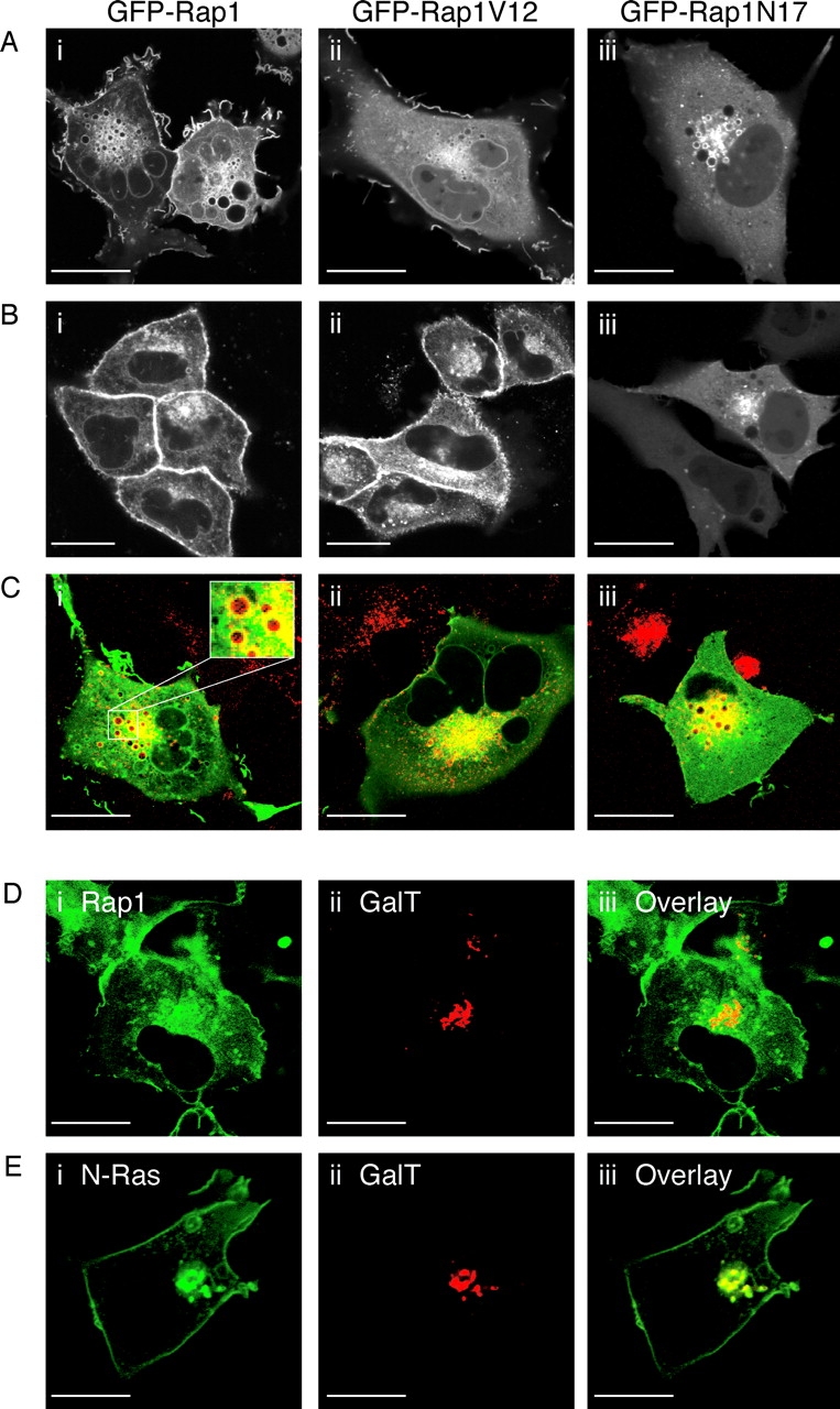
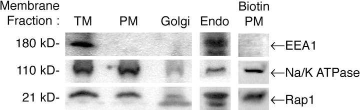
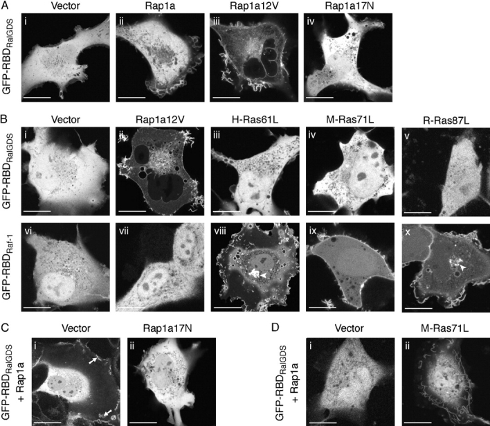
Rap1 and Ras are closely related GTPases that share some effectors but have distinct functions. We studied the subcellular localization of Rap1 and its sites of activation in living cells. Both GFP-tagged Rap1 and endogenous Rap1 were localized to the plasma membrane (PM) and endosomes. The PM association of GFP-Rap1 was dependent on GTP binding, and GFP-Rap1 was rapidly up-regulated on this compartment in response to mitogens, a process blocked by inhibitors of endosome recycling. A novel fluorescent probe for GTP-bound Rap1 revealed that this GTPase was transiently activated only on the PM of both fibroblasts and T cells. Activation on the PM was blocked by inhibitors of endosome recycling. Moreover, inhibition of endosome recycling blocked the ability of Rap1 to promote integrin-mediated adhesion of T cells. Thus, unlike Ras, the membrane localizations of Rap1 are dynamically regulated, and the PM is the principle platform from which Rap1 signaling emanates. These observations may explain some of the biological differences between these GTPases.


Figures

Figure 1.
Figure 1.
Localization of GFP-Rap1 in living cells. COS-1 (A) or MDCK (B) cells were transfected with GFP-Rap1 (i), GFP-Rap1V12 (ii), or GFP-Rap1N17 (iii) and imaged 24 h after transfection with a laser scanning confocal microscope. Results are representative of three independent experiments (>30 cells examined per condition per experiment). (C) COS-1 cells were transfected as in A and incubated at 37°C for 30 min with Texas red–conjugated transferrin. Dual color confocal images were acquired showing colocalization in yellow. Enlarged inset in panel i shows transferrin in the lumen of endosomes decorated with GFP-Rap1. (D and E) COS-1 cells were cotransfected with CFP-GalT and either GFP-Rap1 (D) or GFP-N-Ras (E) and imaged with a laser scanning confocal microscope equipped with the Meta™ system of spectral deconvolution capable of separating CFP from GFP. Bars, 10 μM.
Figure 2.
Figure 2.
Localization of endogenous Rap1. Subcellular fractionation of MDCK cells using sucrose followed by Optiprep gradients or biotinylation cell surface expression assay. Immunoblot analysis for EEA1, Na/K ATPase, and Rap1 was performed on the total membrane fraction (TM) and on membrane fractions highly enriched for the Golgi complex (Golgi), plasma membranes (PM), or endosomes (Endo) obtained by Optiprep, or on PM obtained by biotin affinity purification (Biotin PM). Results are representative of two independent experiments.
Figure 3.
Figure 3.
Growth factor stimulates exocytosis-dependent up-regulation of Rap1 on the PM. (A) COS-1 cells were transfected with GFP-Rap1 wild type without (top and middle) or with cotransfection of dominant-negative Rab11BP (bottom). Cells were serum starved 24 h after transfection, stimulated with EGF in the absence (top and bottom) or presence (middle) of NEM, and imaged before and 5 min after stimulation. Arrow indicates areas of GFP-Rap1 up-regulation on PM ruffles. PM up-regulation of Rap1 was observed in all control cells and validated by measurement of relative fluorescence intensity as described in Materials and methods. In contrast, only 16 ± 9% and 8 ± 8% (mean ± SEM) of NEM-treated and dominant-negative Rab11BP (DN Rab11BP) transfected cells, respectively, showed up-regulation on PM (n = 4; P < 0.0001 for each condition compared with control). (B) COS-1 cells expressing GFP-H-Ras that were serum starved and stimulated as in A showed no change in Golgi apparatus (arrowhead) or PM (arrow) expression. Images shown are representative of seven Z slices acquired to compensate for minimal focal drift. Bars, 10 μM.
Figure 4.
Figure 4.
Rap1 activation in living COS-1 cells. (A) COS-1 cells were transfected with GFP-RBDRalGDS and vector (i), untagged Rap1 wild type (ii), Rap1V12 (iii), or Rap1N17 (iv), and cells were imaged alive 24 h after transfection under conditions of growth in serum. (B) Cos-1 cells were cotransfected with GFP- RBDRalGDS (i–v), GFP–RBDRaf-1 (vi–x), and either vector (i and vi), untagged Rap1V12 (ii and vii), untagged H-Ras61L (iii and viii), untagged M-Ras71L (iv and ix), or untagged R-Ras87L (v and x); serum starved; and imaged as in A. Arrowheads indicate the Golgi apparatus. (C) COS-1 cells were cotransfected with GFP– RBDRalGDS, untagged Rap1 wild type, and either vector (i) or untagged Rap1N17 (ii); grown in serum; and imaged as in A. (D) COS-1 cells were cotransfected with GFP–RBDRalGDS, untagged Rap1 wild type, and either vector (i) or M-Ras71L (ii); serum starved; and imaged as in A. Arrows indicate PM. Bars, 10 μM. Results are representative of three independent experiments (>30 cells examined per condition per experiment).
Figure 5.
Figure 5.
Growth factor–stimulated activation of Rap1 in living cells. (A) COS-1 cells were transfected with GFP-RBDRalGDS, serum starved 24 h after transfection, and imaged alive at the indicated times after stimulation with EGF at 37°C. Endomembrane recruitment of the probe was not observed in any COS-1 cell. PM recruitment of the probe was observed in 60 ± 5% (mean ± SEM) of transfected cells (n = 4). (B) COS-1 or (C) NIH 3T3 cells were cotransfected with GFP-RBDRalGDS and untagged Rap1 wild type, serum starved 24 h after transfection, and imaged as in A. Arrows indicate GFP-RBDRalGDS recruitment to PM. Bars, 10 μM. Endomembrane recruitment of the probe was observed in no COS-1 or NIH-3T3 cell. PM recruitment of the probe was observed in 75 ± 8% and 71 ± 10% (mean ± SEM) of COS-1 and NIH-3T3 cells, respectively (n = 4). (D) COS-1 cells were serum starved, and then stimulated with vehicle or 100 ng/ml EGF for 5 min. Clarified cell lysates were immunoprecipitated with either Sepharose-conjugated GST-RBDRaf-1 and GST-RBDRalGDS (top) or anti-Ras and anti-Rap1 polyclonal antisera (bottom). Immunoprecipitates were immunoblotted with anti-Ras (left) or anti-Rap1 (right) mAbs. Results shown are representative of two independent experiments.
Figure 6.
Figure 6.
Activation of Rap1 at the PM is dependent on exocytosis but not endocytosis. (A) COS-1 cells were transfected with GFP- RBDRalGDS alone (top and middle) or with dominant-negative Rab11BP (DN Rab11BP; bottom), serum starved 24 h after transfection, and stimulated at 37°C with EGF in the absence (top and bottom) or presence (middle) of NEM. Endomembrane recruitment of the probe was observed in no cell. PM recruitment was observed in 63 ± 14%, 25 ± 8%, and 29 ± 10% of control, NEM-treated, and DN Rab11BP transfected cells (mean ± SEM), respectively (n = 4; P < 0.02 for each condition compared with control). (B) COS-1 cells were cotransfected with GFP-RBDRalGDS, untagged Rap1 wild type, and dominant-negative epsin; serum starved; stimulated with EGF in the presence of Texas red–conjugated transferrin; and imaged after 5 min as in A. The dual color image shown in the far right panel was acquired 30 min after stimulation following removal of excess transferrin. Only the untransfected cell to the right accumulated transferrin in endosomes (arrowhead). PM recruitment of the probe was observed in 71 ± 17% (mean ± SEM) of epsin-transfected, EGF-stimulated cells (n = 4). Arrows indicate GFP-RBDRalGDS recruitment to PM. Bars, 10 μM.
Figure 7.
Figure 7.
Up-regulation and activation of Rap1 on PM controls T cell adhesion. (A) Jurkat T cells were transfected with either GFP-Rap1 wild type (i), GFP-Rap1V12 (ii), or GFP-Rap1N17 (iii) and imaged alive 48 h after transfection and growth in serum. (B) Jurkat cells were transfected with GFP-RBDRalGDS and untagged Rap1 wild type (i), Rap1V12 (ii), or Rap1N17 (iii) and imaged as in A. Bars, 2 μM. Results are representative of three independent experiments (>30 cells examined per condition per experiment). (C) Jurkat cells were transfected with GFP-Rap1, serum starved, and treated with or without anti-CD3 antibodies. Cells were scored for unambiguous PM expression and the percentage of such cells before and after cross-linking TCRs was plotted (mean ± SEM, n = 4). Representative cells before (left) and after (right) anti-CD3 treatment are shown as insets. Bars, 2 µM. (D) Jurkat cells were transfected as indicated and plated on fibronectin-coated multi-well plates 48 h later. (E) Jurkat cells were transfected as indicated and plated on ICAM-1–coated multi-well plates 48 h later with or without treatment with anti-CD3 antibodies. Cells per well (in duplicate) bound to fibronectin (D) or ICAM-1 (E) after 1 h were counted and presented as the mean ± SEM (n = 4).

Similar articles

Cited by

References

    1. Altschuler, D.L., and F. Ribeiro-Neto. 1998. Mitogenic and oncogenic properties of the small G protein Rap1b. Proc. Natl. Acad. Sci. USA. 95:7475–7479. - PMC - PubMed
    1. Beranger, F., B. Goud, A. Tavitian, and J. de Gunzburg. 1991. Association of the Ras-antagonistic Rap1/Krev-1 proteins with the Golgi complex. Proc. Natl. Acad. Sci. USA. 88:1606–1610. - PMC - PubMed
    1. Berger, G., R. Quarck, D. Tenza, S. Levy-Toledano, J. de Gunzburg, and E.M. Cramer. 1994. Ultrastructural localization of the small GTP-binding protein Rap1 in human platelets and megakaryocytes. Br. J. Haematol. 88:372–382. - PubMed
    1. Bos, J.L. 1998. All in the family? New insights and questions regarding interconnectivity of Ras, Rap1 and Ral. EMBO J. 17:6776–6782. - PMC - PubMed
    1. Bos, J.L., J. de Rooij, and K.A. Reedquist. 2001. Rap1 signalling: adhering to new models. Nat. Rev. Mol. Cell Biol. 2:369–377. - PubMed

Publication types

MeSH terms