. 2004 Feb 10;101(6):1589-94.
doi: 10.1073/pnas.0305802101. Epub 2004 Feb 2.

Rapid modulation of osteoblast ion channel responses by 1alpha,25(OH)2-vitamin D3 requires the presence of a functional vitamin D nuclear receptor

Affiliations

Rapid modulation of osteoblast ion channel responses by 1alpha,25(OH)2-vitamin D3 requires the presence of a functional vitamin D nuclear receptor

Laura P Zanello et al. Proc Natl Acad Sci U S A. .

Abstract

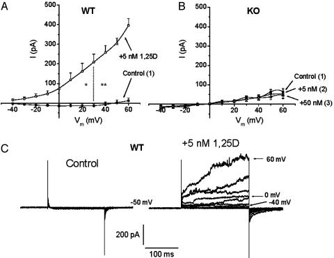
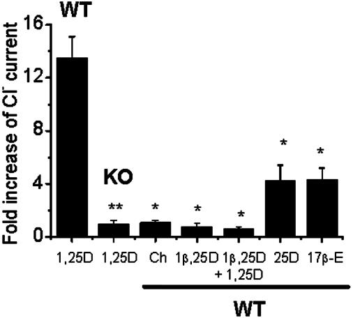
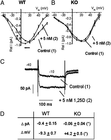
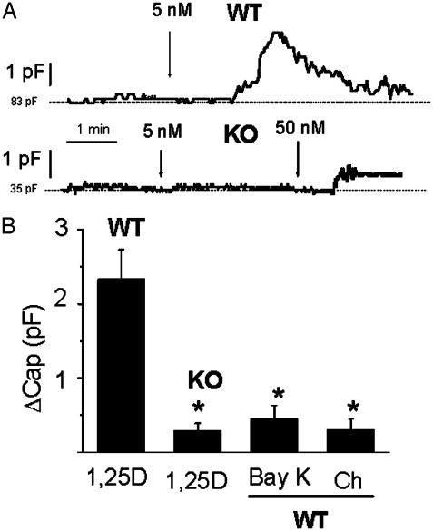
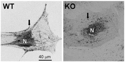
1alpha,25(OH)(2)-Vitamin D(3) (1,25D) modulates osteoblast gene expression of bone matrix proteins via a nuclear vitamin D receptor (VDR) and also modifies the electrical state of the plasma membrane through rapid nongenomic mechanisms still not fully understood. The physiological significance of 1,25D membrane-initiated effects remains unclear. To elucidate whether the VDR is required for 1,25D-promoted electrical responses, we studied 1,25D modulation of ion channel activities in calvarial osteoblasts isolated from VDR knockout (KO) and WT mice. At depolarizing potentials, Cl(-) currents were significantly potentiated (13.5 +/- 1.6-fold increase, n = 12) by 5 nM 1,25D in VDR WT but not in KO (0.96 +/- 0.3 fold increase, n = 11) osteoblasts. L-type Ca(2+) currents significantly shift their peak activation by -9.3 +/- 0.7 mV (n = 10) in the presence of 5 nM 1,25D in VDR WT but not in KO cells, thus facilitating Ca(2+) influx. Furthermore, we found that 1,25D significantly increased whole-cell capacitance in VDR WT (DeltaCap = 2.3 +/- 0.4 pF, n = 8) but not in KO osteoblasts (DeltaCap = 0.3 +/- 0.1 pF, n = 8); this corresponds to a rapid (1-2 min) fusion in WT of 71 +/- 33 versus in KO only 9 +/- 6 individual secretory granules. We conclude that, in calvarial osteoblasts, 1,25D modulates ion channel activities only in cells with a functional VDR and that this effect is coupled to exocytosis. This is a demonstration of the requirement of a functional classic steroid receptor for the rapid hormonal modulation of electric currents linked to secretory activities in a target cell.


Figures

Fig. 1.
Fig. 1.
1,25D potentiation of Cl- currents in VDR WT but not in KO osteoblasts. Current to voltage (I/Vm) relations obtained for Cl- channel activities from VDR WT (A) and KO (B) osteoblasts, before (filled circles) and after the addition of 5 nM (open circles) and 50 nM (open triangles) 1,25D to the bath. To allow for volume-sensitive Cl- currents to reach a stable amplitude value, current amplitudes (I, pA) were measured 5–10 min after obtaining the whole-cell configuration (control curves) and 5–10 min after the addition of the hormone to the bath. The current was measured at the peak of maximal activation of outward currents elicited by a series of 200-ms depolarizing voltage steps between -40 and 60 mV from a holding potential of -50 mV, applied every second. Values represent the mean ± SEM of n = 12 WT and n = 11 KO osteoblasts. Numbers in parentheses indicate the sequential order of recordings obtained from the same single cell. (C) Typical raw currents recorded from a single VDR WT osteoblast. Recording solutions are described in Materials and Methods.
Fig. 2.
Fig. 2.
Cl- current potentiation in VDR WT osteoblasts is selective for 1,25D. Average fold increase of outward Cl- currents measured at 60 mV, 5–10 min after the addition of 5 nM 1,25D (n = 12 VDR WT and n = 11 KO osteoblasts), 0.1–1 μM cholesterol (Ch, n = 5), 10 nM of the synthetic analog 1β,25D by itself (n = 3) or followed by the addition of 5 μM 1,25D (n = 3), 5 nM the natural metabolite 25D (n = 4), and 0.1–1 μM17β-estradiol (17β-E, n = 4) to the bath. The magnitude of the response was compared to the fold increase obtained with 5 nM 1,25D on VDR WT osteoblasts. Values represent the mean ± SEM. *, P < 0.05; **, P < 0.01.
Fig. 3.
Fig. 3.
Effects of 1,25D on L-type Ca2+ channels in VDR WT and KO osteoblasts. I/Vm relations obtained for Ca2+ channel activities from VDR WT (A) and KO (B) osteoblasts, before (filled circles) and 5 min after (open circles) the addition of 5 nM 1,25D to the bath. Rel I (pA) represents the relative current value obtained for each series of 200-ms depolarizing voltage steps in relation to the maximal I (pA) value obtained for each single cell. Voltage steps were applied every second between 40 and 60 mV, from a holding potential of -40 mV. Values represent the mean ± SEM of n = 10 WT and n = 6 KO osteoblasts. Numbers in parentheses indicate the sequential order of recordings obtained from the same single cell. (C) Raw data for the typical potentiation of inward Ba2+ currents by 1,25D obtained at -10 mV from a single VDR WT osteoblast. (D) Summary of the values obtained for current amplitude increments (ΔpA) at -10 mV and shifts in I/Vm relationships (ΔmV) due to the addition of 5 nM 1,25D to VDR WT (n = 10) and KO (n = 6) osteoblasts. Data (average ± SEM) from WT and KO cells were statistically different (*, P < 0.50). Recording solutions are described in Materials and Methods.
Fig. 4.
Fig. 4.
1,25D promoted an increase in whole-cell capacitance in VDR WT but not in KO osteoblasts. (A) Continuous recordings of whole-cell capacitance values obtained from a VDR WT and a KO osteoblast during the addition of 5 nM (WT) and 5–50 nM (KO) 1,25D to the bath. Cell capacitance was automatically measured every 1 sec. (B) Increments in whole-cell capacitance value (ΔCap) measured after the addition of 5 nM 1,25D (n = 8 WT and n = 8 KO cells), 1 μM (±)-Bay K8644 (n = 4), and 1 μM cholesterol (Ch, n = 3). ΔCap values (average ± SEM) were calculated as the difference between the peak of maximal capacitance value achieved after the addition of the agent and the initial basal line (dashed line) and were statistically compared to the increase produced by 5 nM 1,25D in VDR WT osteoblasts.*, P < 0.05.
Fig. 5.
Fig. 5.
Laser scanning confocal images obtained from a VDR WT and a VDR KO osteoblast in culture for 3 weeks. Secretory granules (arrows) labeled with 1 μM quinacrine (see Materials and Methods) distribute abundantly over the cytoplasm and adjacent to the cell membrane in a VDR WT osteoblast but are scarce in the cytoplasm and virtually absent from the cell periphery in a KO osteoblast. These results are typical of ≈50 VDR WT and KO cells studied.

References

    1. Caffrey, J. M. & Farach-Carson, M. C. (1989) J. Biol. Chem. 264, 20265-20274. - PubMed
    1. Zanello, L. P. & Norman, A. W. (1997) J. Biol. Chem. 272, 22617-22622. - PubMed
    1. Song, X., Bishop, J. E., Okamura, W. H. & Norman, A. W. (1998) Endocrinology 139, 457-465. - PubMed
    1. Bouillon, R., Okamura, W. H. & Norman, A. W. (1995) Endocr. Rev. 16, 200-257. - PubMed
    1. Kitanaka, S., Takeyama, K., Murayama, A., Sato, T., Okumura, K., Nogami, M., Hasegawa, Y., Niimi, H., Yanagisawa, J., Tanaka, T., et al. (1998) N. Engl. J. Med. 338, 653-661. - PubMed

Publication types

LinkOut - more resources