. 2004 Feb 10;101(6):1578-83.
doi: 10.1073/pnas.0303274101. Epub 2004 Feb 2.

Ca2+/calmodulin transfers the membrane-proximal lipid-binding domain of the v-SNARE synaptobrevin from cis to trans bilayers

Affiliations

Ca2+/calmodulin transfers the membrane-proximal lipid-binding domain of the v-SNARE synaptobrevin from cis to trans bilayers

Luc de Haro et al. Proc Natl Acad Sci U S A. .

Abstract

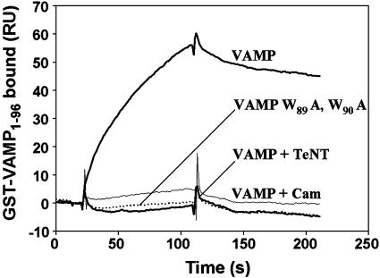
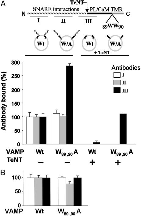
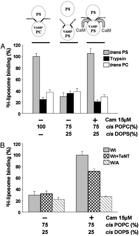
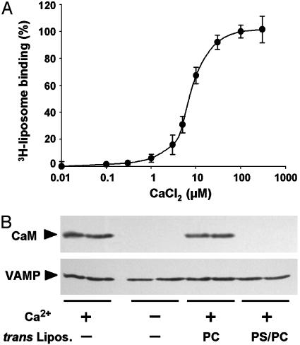
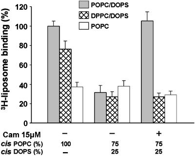
Soluble N-ethylmaleimide-sensitive fusion protein attachment protein receptor (SNARE) protein interactions at the synaptic vesicle/plasma membrane interface play an essential role in neurotransmitter release. The membrane-proximal region (amino acids 77-90) of the v-SNARE vesicle-associated membrane protein 2 (VAMP 2, synaptobrevin) binds acidic phospholipids or Ca(2+)/calmodulin in a mutually exclusive manner, processes that are required for Ca(2+)-dependent exocytosis. To address the mechanisms involved, we asked whether this region of VAMP can interact with cis (outer vesicle leaflet) and/or trans (inner plasma membrane leaflet) lipids. To evaluate cis lipid binding, recombinant VAMP was reconstituted into liposomes and accessibility to site-directed antibodies was probed by surface plasmon resonance. Data indicated that the membrane-proximal domain of VAMP dips into the cis lipid bilayer, sequestering epitopes between the tetanus toxin cleavage site and the membrane anchor. These epitopes were unmasked by VAMP double mutation W89A, W90A, which abolishes lipid interactions. To evaluate trans lipid binding, VAMP was reconstituted in cis liposomes, which were then immobilized on beads. The ability of VAMP to capture protein-free (3)H-labeled trans liposomes was then measured. When cis lipid interactions were eliminated by omitting negatively charged lipids, trans lipid binding to VAMP was revealed. In contrast, when cis and trans liposomes both contained acidic headgroups (i.e., approximating physiological conditions), cis lipid interactions totally occluded trans lipid binding. In these conditions Ca(2+)/calmodulin displaced cis inhibition, transferring the lipid-binding domain of VAMP from the cis to the trans bilayer. Our results suggest that calmodulin acts as a unidirectional Ca(2+)-activated shuttle that docks the juxtamembrane portion of the v-SNARE in the target membrane to prepare fusion.


Figures

Fig. 1.
Fig. 1.
Binding of the membrane-proximal domain of VAMP to acidic phospholipids. Liposomes (25% DOPS/75% POPC) were immobilized on a hydrophobic sensor chip of an SPR (Biacore) apparatus. GST-VAMP1–96 (50 nM, thick upper trace) was injected in a running buffer containing 5 mM Hepes/NaOH, 100 mM NaCl, and 1 mM CaCl2, pH 7.4. Binding was inhibited by pretreatment with TeNT, 10 μg/ml, 1 h at 37°C, which cleaves the Q76–F77 peptide bond (thick lower trace), by introduction of the W89A, W90A mutation (dotted trace), and by coinjection of 10 μM calmodulin (thin trace).
Fig. 2.
Fig. 2.
Cis lipid binding masks epitopes in the membrane-proximal domain of VAMP. Upper schema illustrates VAMP domains recognized by antibodies I, II, and III. The membrane-proximal region (black) is flanked N-terminally by the TeNT cleavage site (Q76–F77) and C-terminally by the transmembrane anchor (TMR). The mutation W89A, W90A inhibits lipid binding. PL, phospholipid; CaM, calmodulin. Lower schema represents the four VAMP liposome preparations. (A) Full-length recombinant wild type (Wt) or W89A, W90A mutant (W/A) VAMP was reconstituted into 25% DOPS/75% POPC liposomes (Left), and aliquots of each were treated with TeNT (Right). The four resulting liposome pools were immobilized on the sensor chip of an SPR apparatus and the binding of antibodies I (white bars), II (gray bars), and III (black bars) to each individual group was assayed. The data are presented as four groups of three bars. Bound antibody = mass (RU) of antibody bound/mass (RU) of VAMP liposome immobilized. Binding of each antibody to wild-type VAMP liposomes was normalized to 100%. Antibody III binding was corrected to account for direct effects of the W89A, W90A mutation (Fig. 6C). After TeNT treatment, binding of antibodies I and II was not detectable. Results are the means ± SD of three independent experiments, each in triplicate. (B) As in A Left, except that VAMP was reconstituted into 100% POPC. Results are the means ± SD of an experiment in quadruplicate.
Fig. 3.
Fig. 3.
Ca2+/calmodulin transfers the lipid-binding domain of VAMP from cis to trans bilayers. (A) The schema at the top represents the three assay conditions that correspond to the gray bars in A, (black and white bars represent negative controls). Wild-type VAMP was reconstituted into liposomes of the indicated lipid composition. Cis VAMP liposomes were immobilized on beads and incubated with trans 3H-liposomes containing acidic headgroups (25% DOPS/75% POPC, gray bars) in the presence of 1 mM Ca2+ and in the presence or absence of calmodulin (CaM). After washing by centrifugation, bound radioactivity was measured. Nonspecific binding was evaluated with trypsin-treated cis liposomes (black bars) and 100% POPC trans 3H-liposomes (white bars). Trans 3H-liposome binding to VAMP reconstituted in 100% POPC cis liposomes was set at 100%. Results are the means ± SD of six independent experiments, each in triplicate. (B) Wild-type VAMP (gray and cross-hatched bars) or W89A, W90A VAMP (hatched bars) were reconstituted into cis liposomes (25% DOPS/75% POPC) and immobilized on beads. Wild-type VAMP liposomes were preincubated in the absence (gray bars) or presence (cross-hatched bars) of TeNT. Immobilized VAMP liposomes on beads were then incubated with trans 3H-liposomes (25% DOPS/75% POPC) in the presence and absence of calmodulin and processed as in A. 3H-liposome binding to native VAMP liposomes in the presence of calmodulin was set at 100%. Results are the means ± SD of two independent experiments, each in triplicate.
Fig. 4.
Fig. 4.
Ca2+/calmodulin binding to cis liposomes mediates transfer of the lipid-binding domain of VAMP. (A) Wild-type VAMP liposomes (25% DOPS/75% POPC) immobilized on beads were incubated with 15 μM calmodulin and trans 3H-liposomes (25% DOPS/75% POPC) in the presence of a Ca2+/EGTA buffer to yield the indicated free Ca2+ concentrations. 3H-liposome binding was measured as in Fig. 3. Results are the means ± SD of two independent experiments, each in triplicate. (B) Wild-type VAMP liposomes (as in A) were incubated with 15 μM calmodulin in the presence or absence of 150 μMCa2+ and trans liposomes (100% POPC or 25% DOPS/75% POPC) as indicated. After washing, calmodulin binding and VAMP content were monitored by Western blotting. Each experimental condition was studied in duplicate, and the products were run on adjacent gel lanes. Results are representative of two independent experiments.
Fig. 5.
Fig. 5.
Unidirectional Ca2+/calmodulin-dependent transfer of the lipid-binding domain of VAMP requires fluid-phase trans bilayers. Wild-type VAMP was reconstituted into liposomes with the indicated cis lipid composition. Binding was assayed as in Fig. 3A using trans 3H-liposomes in fluid phase (25% DOPS/75% POPC, gray bars), gel phase (25% DOPS/75% DPPC, cross-hatched bars), or fluid phase lacking acidic headgroups (100% POPC, white bars). Results are the means ± SD of three independent experiments, each in triplicate.

References

    1. Sollner, T., Whiteheart, S. W., Brunner, M., Erdjument-Bromage, H., Geromanos, S., Tempst, P. & Rothman, J. E. (1993) Nature 362, 318-324. - PubMed
    1. McNew, J. A., Parlati, F., Fukuda, R., Johnston, R. J., Paz, K., Paumet, F., Sollner, T. H. & Rothman, J. E. (2000) Nature 407, 153-159. - PubMed
    1. Weber, T., Zemelman, B. V., McNew, J. A., Westermann, B., Gmachl, M., Parlati, F., Sollner, T. H. & Rothman, J. E. (1998) Cell 92, 759-772. - PubMed
    1. Jahn, R. & Südhof, T. C. (1999) Annu. Rev. Biochem. 68, 863-911. - PubMed
    1. Mayer, A. (1999) Curr. Opin. Cell Biol. 11, 447-452. - PubMed

Publication types

LinkOut - more resources