. 2004 Feb 2;32(2):700-9.
doi: 10.1093/nar/gkh231. Print 2004.

The Trypanosoma brucei spliced leader RNA and rRNA gene promoters have interchangeable TbSNAP50-binding elements

Affiliations

The Trypanosoma brucei spliced leader RNA and rRNA gene promoters have interchangeable TbSNAP50-binding elements

Bernd Schimanski et al. Nucleic Acids Res. .

Abstract

In the protist parasite Trypanosoma brucei, the small nuclear spliced leader (SL) RNA and the large rRNAs are key molecules for mRNA maturation and protein synthesis, respectively. The SL RNA gene (SLRNA) promoter recruits RNA polymerase II and consists of a bipartite upstream sequence element (USE) and an element close to the transcription initiation site. Here, we analyzed the distal part of the ribosomal (RRNA) promoter and identified two sequence blocks which, in reverse orientation, closely resemble the SLRNA USE by both sequence and spacing. A detailed mutational analysis revealed that the ribosomal (r)USE is essential for efficient RRNA transcription in vivo and that it functions in an orientation-dependent manner. Moreover, we showed that USE and rUSE are functionally interchangeable and that rUSE stably interacted with an essential factor of SLRNA transcription. Finally, we demonstrated that the T.brucei homolog of the recently characterized transcription factor p57 of the related organism Leptomonas seymouri specifically bound to USE and rUSE. Since p57 and its T.brucei counterpart are homologous to SNAP50, a component of the human small nuclear RNA gene activation protein complex (SNAPc), both SLRNA and RRNA transcription in T.brucei may depend on a SNAPc-like transcription factor.


Figures

Figure 1
Figure 1
Similar sequence elements are present in T.brucei RRNA and SLRNA promoters. (A) The double-stranded sequence of the RRNA promoter from position –240 to position –197 relative to the transcription initiation site is aligned with the reversed sense strand of the SLRNA promoter spanning positions –39 to –82. Identical nucleotides in the antisense strand of the RRNA promoter and the sense strand of the SLRNA promoter are drawn in bold letters and shaded in gray. The two essential SLRNA promoter elements USE1 and USE2 are underlined. (B) Schematic outline to scale of RRNA and SLRNA promoters. Promoter elements are represented as boxes and the transcription initiation sites by flags. RRNA promoter domain IV consists of two sequence blocks which were designated as rUSE1 and rUSE2 and, as their SLRNA counterparts USE1 and USE2, are drawn as black boxes. The arrows indicate the opposite orientation of USE and rUSE in their respective promoters. The stippled line of the box representing RRNA promoter domain III indicates that promoter sequences have not been mapped in this domain.
Figure 2
Figure 2
In vitro competition of Rib-trm and SLins19 transcription with linear RRNA promoter fragments. (A) Schematic outline to scale of the RRNA promoter (see legend of Fig. 1) and of the competitor fragments RRNA –257/–3, RRNA –162/–3 and RRNA –257/–162. (B) Transcription competition analysis. Template constructs Rib-trm or SLins19 were transcribed in vitro in the presence of linear DNA competitor fragments. As competitors, a 222 bp non-specific linear DNA fragment (nonspec) or RRNA promoter fragments were used in a 10-fold molar excess to template DNA. Rib-trm and SLins19 transcripts were detected by primer extension of total RNA prepared from transcription reactions with 5′-end-labeled oligonucleotides Tag_PE and SLtag, respectively. In control reactions, endogenous U2 snRNA was detected in the same RNA preparations by primer extension using the 5′-end-labeled oligonucleotide U2k. Primer extension products were separated on 6% polyacrylamide–50% urea gels and visualized by autoradiography. Arrows on the right point to primer extension signals of Rib-trm, SLins19 and endogenous U2 (end U2) RNAs. M, marker (MspI- digested pBR322); lengths of marker fragments are indicated on the left.
Figure 3
Figure 3
rUSE and USE stably interact with a trans-activating SLRNA transcription factor. (A) Schematic outline of RRNA –257/–162 and SLRNA –126/–18 competitor fragments and their mutated versions (r)USE1-mut and (r)USE2-mut. (r)USE1 and (r)USE2 are represented by black boxes and mutated regions by gray boxes. Mutation of rUSE1 and USE1 comprised the substitution of the inner 10 bp by the sequence 5′-TGACATATGA-3′, whereas rUSE2 and USE2 were completely replaced by the sequence 5′-CTTGACATATGC-3′. These sequences relate to the orientation of rUSE and USE as indicated by arrows. (B) Competition of SLins19 transcription. In vitro transcription reactions were carried out in the presence of a 10-fold molar excess of linear DNA competitor fragments. SLins19 transcription signals and the endogenous U2 snRNA control signals (end U2) were obtained by primer extension assays of total RNA with 5′-end-labeled oligonucleotides. The primer extension products were separated by denaturing gel electrophoresis and visualized by autoradiography. M, marker MspI-digested pBR322.
Figure 4
Figure 4
Block substitution analysis of RRNA promoter domain IV. (A) Depicted are sequences of the wild-type RRNA promoter domain IV in the construct RibCAT and of the block substitutions in the mutational constructs RibCAT1–6. Numbering is relative to the transcription initiation site, and the sequences of rUSE1 and rUSE2 are underlined. Unchanged nucleotides are indicated by dots. (B) Transient transfection analysis of promoter mutations. The wild-type RibCAT (WT) and the mutational constructs RibCAT1–6 were transfected into procyclic T.brucei cells together with the control plasmid TU2∇81 carrying an oligonucleotide-tagged U2 snRNA gene. At 16 h after transfection, total RNA was prepared from transfected cells and analyzed by primer extension with the 5′-end-labeled oligonucleotides CAT5 and U2-BTAG which are complementary to the CAT-coding region and the TU2∇81 tag sequence, respectively. Primer extension products were separated on a 6% polyacrylamide–50% urea gel and visualized by autoradiography. On the left, positions of MspI-digested pBR322 marker fragments are indicated, and on the right, arrows point to the primer extension products of CAT mRNA and TU2∇81 snRNA (U2). (C) Primer extension signals of three independent experiments were quantified by densitometry. The CAT signal strengths were standardized with TU2∇81 signals and the signal strength of wild-type RibCAT was set to 100%. Means and standard deviations are graphically depicted.
Figure 5
Figure 5
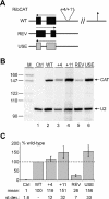
Manipulation of RRNA promoter domain IV. (A) Schematic outline of mutant constructs. In plasmids RibCAT +4 and RibCAT +11, rUSE was moved further upstream of the transcription initiation site by 4 and 11 bp, respectively. In construct RibCAT-REV, rUSE was replaced by its reverse complement and in construct RibCAT-USE by the corresponding sequence of the SLRNA promoter. (B) In transient expression experiments, RibCAT constructs were co-transfected with the control plasmid TU2∇81 into procyclic cells. In a control experiment (Ctrl), only the TU2∇81 plasmid was transfected. CAT and TU2∇81 expression was analyzed as described in Figure 4 by primer extension of total RNA prepared from transfected cells. M, marker MspI-digested pBR322; lengths of marker fragments are indicated on the left. Arrows on the right point to primer extension products of CAT mRNA and TU2∇81 snRNA (U2). (C) Densitometric analysis of CAT and TU2∇81 signal strengths in three independent experiments. The standardized RibCAT (WT) signal strength was set to 100. Means and standard deviations (st.dev.) obtained with mutant constructs are given in graphic and numeric form.
Figure 6
Figure 6
rUSE can partially replace USE function in the SLRNA promoter. Standard in vitro transcription reactions were carried out with SLins19 constructs carrying the SLRNA wild-type promoter or derivatives with linker scanner mutations in USE1 (LS –71/–62) or USE2 (LS –53/–42). In construct SLins19-rUSE (rUSE), the complete USE sequence from position –71 to position –42 was replaced by the corresponding sequence of rUSE. As a control, endogenous U2 snRNA (end U2) was detected in each reaction by primer extension of oligonucleotide U2f. Below each lane, means and standard deviations of standardized transcription signal strengths derived from six independent experiments are given relative to the wild-type SLins19 signal which was set to 100. The arrow on the right points to the specific primer extension signal of SLins19 RNA. The band below the main signal appears in some transcription extracts and may be caused by in vitro methylation of 5′-terminal SLins19 RNA nucleotides which is known to terminate primer extension signals prematurely. On the left, sizes of pBR322 MspI marker fragments are indicated.
Figure 7
Figure 7
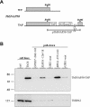
TbSNAP50 binds to USE and rUSE. (A) Illustration of the two TbSNAP50 alleles (not to scale) in the procyclic cell line TbC8. Shown are the unaltered wild-type allele (WT) and the modified allele in which the TAP sequence was fused 3′ terminally to the TbSNAP50 coding region by targeted insertion of the BstBI-linearized construct pTbSNAP50-TAP (TAP). As a selection marker, the construct contained the neomycin phosphotransferase gene (neo) flanked by HSP70 genes 2 and 3 (H23) and βα tubulin (T) intergenic regions. (B) Immunoblot analysis of transcription extract prepared from TbC8 or control cells (ctrl), and of proteins bound to immobilized DNA fragments (pull-down). The latter comprised procyclin GPEET promoter domain IV (GPEET –246/–162), the wild-type SLRNA promoter upstream region (SLRNA –126/–18) and a corresponding fragment carrying a mutation in USE1 (USE1-mut) as well as the wild-type RRNA promoter domain IV (RRNA –257/–162) and an equivalent fragment with a mutation in rUSE1 (rUSE1-mut). The same blot was analyzed with the PAP reagent specific for the protein A epitopes within the TAP tag (TbSNAP50-TAP) and with a polyclonal antibody directed against TbRPA2, the second largest subunit of RNA pol I.

Similar articles

Cited by

References

    1. Günzl A. (2003) Transcription. In Marr,J.J., Nielsen,T.W. and Komuniecki,R.W. (eds), Molecular Medical Parasitology. Academic Press, London, UK, pp. 47–65.
    1. Vanhamme L., Pays,A., Tebabi,P., Alexandre,S. and Pays,E. (1995) Specific binding of proteins to the noncoding strand of a crucial element of the variant surface glycoprotein, procyclin and ribosomal promoters of Trypanosoma brucei. Mol. Cell. Biol., 15, 5598–5606. - PMC - PubMed
    1. Pham V.P., Qi,C.C. and Gottesdiener,K.M. (1996) A detailed mutational analysis of the VSG gene expression site promoter. Mol. Biochem. Parasitol., 75, 241–254. - PubMed
    1. Ginger M.L., Blundell,P.A., Lewis,A.M., Browitt,A., Günzl,A. and Barry,J.D. (2002) Ex vivo and in vitro identification of a consensus promoter for VSG genes expressed by metacyclic-stage trypanosomes in the tsetse fly. Eukaryot. Cell, 1, 1000–1009. - PMC - PubMed
    1. Sherman D.R., Janz,L., Hug,M. and Clayton,C. (1991) Anatomy of the PARP gene promoter of Trypanosoma brucei. EMBO J., 10, 3379–3386. - PMC - PubMed

Publication types

MeSH terms

Associated data