

Lymphatic density and metastatic spread in human malignant melanoma
- PMID: 14760386
- PMCID: PMC2409610
- DOI: 10.1038/sj.bjc.6601571
Lymphatic density and metastatic spread in human malignant melanoma
Abstract
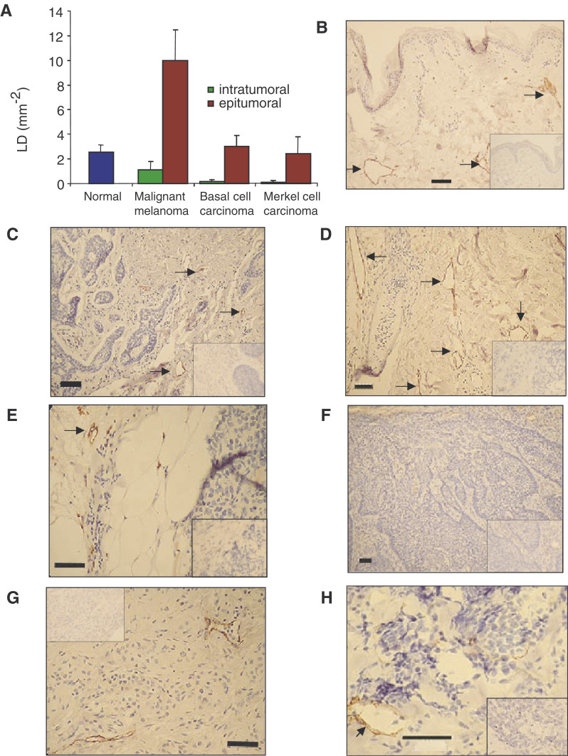
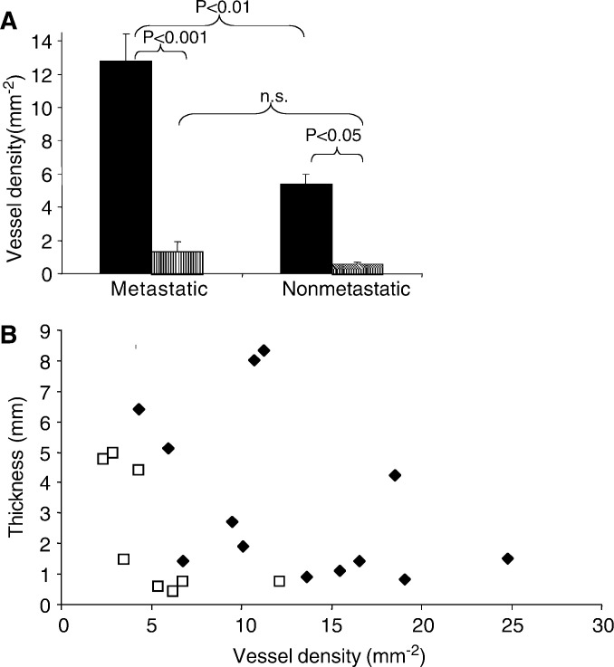
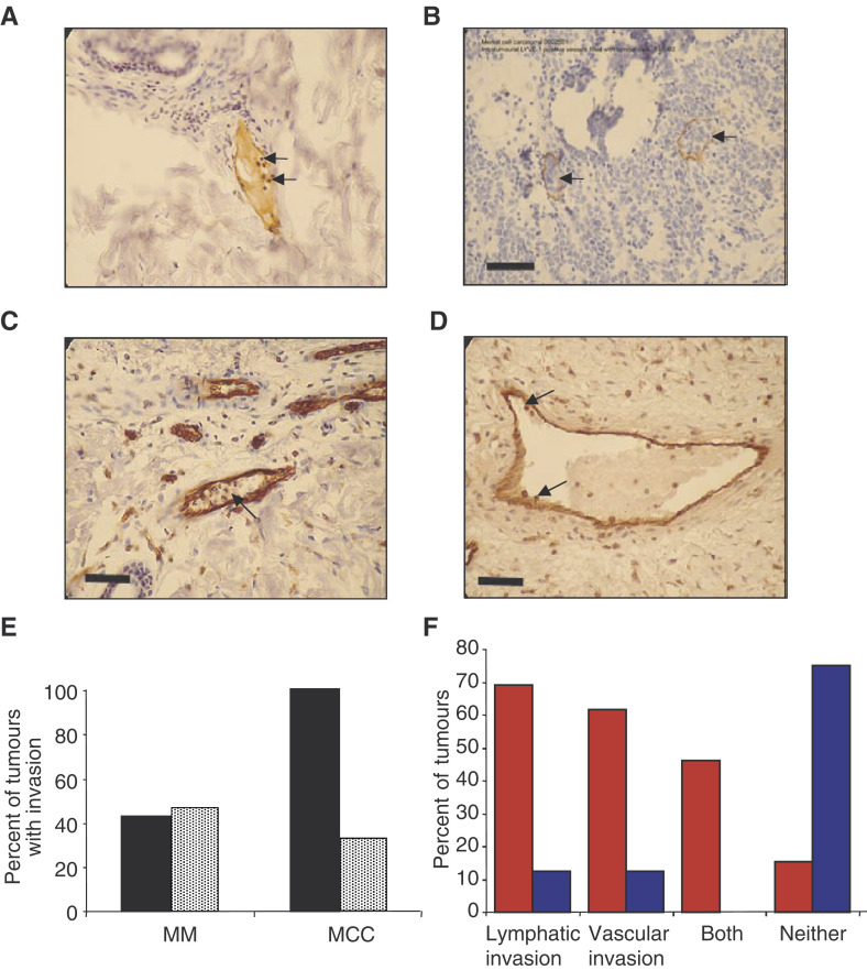
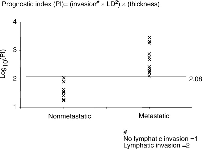
Malignant melanoma (MM), the most common cause of skin cancer deaths, metastasises to regional lymph nodes. In animal models of other cancers, lymphatic growth is associated with metastasis. To assess if lymphatic density (LD) was increased in human MM, and its association with metastasis, we measured LD inside and around archival MM samples (MM, n=21), and compared them with normal dermis (n=11), basal cell carcinoma (BCC, n=6) and Merkel cell carcinoma (MCC), a skin tumour thought to metastasise through a vascular route (MCC, n=6). Lymphatic capillary density (mm(-2)), as determined by immunohistochemical staining with the lymphatic specific marker LYVE-1, was significantly increased around MM (10.0+/-2.5 mm(-2)) compared with normal dermis (2.4+/-0.9 mm(-2)), BCC (3.0+/-0.9 mm(-2)) and MCC (2.4+/-1.4 mm(-2)) (P<0.0001). There was a small decrease in LD inside MM (1.1+/-0.7 mm(-2)) compared with normal dermis, but a highly significant decrease in BCC (0.14+/-0.13) and MCC (0.12+/-2.4) (P<0.01 Kruskal-Wallis). Astonishingly, LD discriminated between melanomas that subsequently metastasised (12.8+/-1.6 mm(-2)) and those that did not (5.4+/-1.1 mm(-2), P<0.01, Mann-Whitney). Lymphatic invasion by tumour cells was seen mainly in MM that metastasised (70% compared with 12% not metastasising, P<0.05 Fisher's Exact test). The results show that LD was increased around MMs, and that LD and tumour cell invasion of lymphatics may help to predict metastasis. To this end, a prognostic index was calculated using LD, lymphatic invasion and thickness that clearly discriminated metastatic from nonmetastatic tumours.
Figures
Comment in
-
Does increased lymphatic density contribute to fast drainage and metastatic spread to sentinel lymph nodes in melanoma?Br J Cancer. 2004 Jul 5;91(1):193. doi: 10.1038/sj.bjc.6601917. Br J Cancer. 2004. PMID: 15187997 Free PMC article. No abstract available.
-
Lymphatic vessel density and prognosis in cutaneous melanoma.Br J Cancer. 2004 Sep 13;91(6):1224-5. doi: 10.1038/sj.bjc.6602122. Br J Cancer. 2004. PMID: 15316566 Free PMC article. No abstract available.
References
-
- Achen MG, Williams RA, Minekus MP, Thornton GE, Stenvers K, Rogers PA, Lederman F, Roufail S, Stacker SA (2001) Localization of vascular endothelial growth factor-D in malignant melanoma suggests a role in tumour angiogenesis. J Pathol 193: 147–154 - PubMed
-
- Baxter LT, Jain RK (1989) Transport of fluid and macromolecules in tumors. I. Role of interstitial pressure and convection. Microvasc Res 37: 77–104 - PubMed
-
- Beasley NJ, Prevo R, Banerji S, Leek RD, Moore J, van Trappen P, Cox G, Harris AL, Jackson DG (2002) Intratumoral lymphangiogenesis and lymph node metastasis in head and neck cancer. Cancer Res 62: 1315–1320 - PubMed
-
- Birner P, Schindl M, Obermair A, Breitenecker G, Kowalski H, Oberhuber G (2001) Lymphatic microvessel density as a novel prognostic factor in early-stage invasive cervical cancer. Int J Cancer 95: 29–33 - PubMed
Publication types
MeSH terms
Grants and funding
LinkOut - more resources
Full Text Sources
Other Literature Sources
Medical
Research Materials
Miscellaneous
