. 2004 Feb;186(4):968-77.
doi: 10.1128/JB.186.4.968-977.2004.

Architecture of the Bacteroides cellulosolvens cellulosome: description of a cell surface-anchoring scaffoldin and a family 48 cellulase

Affiliations

Architecture of the Bacteroides cellulosolvens cellulosome: description of a cell surface-anchoring scaffoldin and a family 48 cellulase

Qi Xu et al. J Bacteriol. 2004 Feb.

Abstract

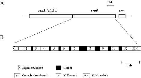
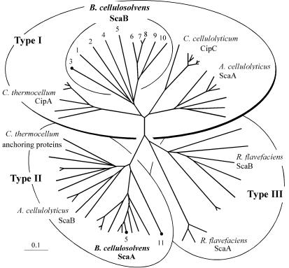
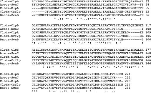
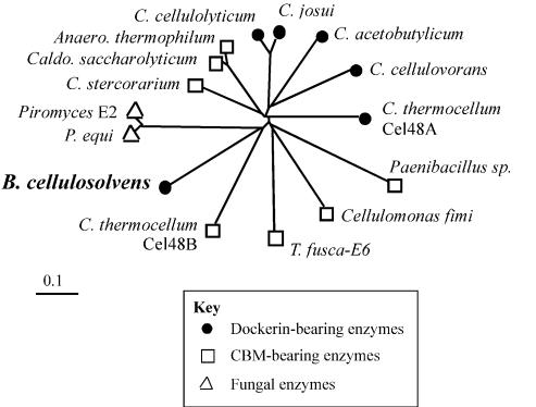
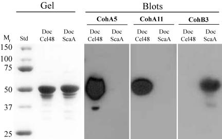
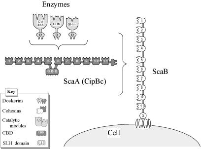
A large gene downstream of the primary Bacteroides cellulosolvens cellulosomal scaffoldin (cipBc, now renamed scaA) was sequenced. The gene, termed scaB, contained an N-terminal leader peptide followed by 10 type I cohesins, an "X" domain of unknown structure and function, and a C-terminal S-layer homology (SLH) surface-anchoring module. In addition, a previously identified gene in a different part of the genome, encoding for a dockerin-borne family 48 cellulosomal glycoside hydrolase (Cel48), was sequenced completely, and a putative cellulosome-related family 9 glycosyl hydrolase was detected. Recombinant fusion proteins, comprising dockerins derived from either the ScaA scaffoldin or Cel48, were overexpressed. Their interaction with ScaA and ScaB cohesins was examined by immunoassay. The results indicated that the ScaB type I cohesin of the new anchoring protein binds selectively to the ScaA dockerin, whereas the Cel48 dockerin binds specifically to the type II ScaA cohesin 5. Thus, by virtue of the 11 type II ScaA cohesins and the 10 type I ScaB cohesins, the relatively simple two-component cellulosome-integrating complex would potentially incorporate 110 enzyme molecules onto the cell surface via the ScaB SLH module. Compared to previously described cellulosome systems, the apparent roles of the B. cellulosolvens cohesins are reversed, in that the type II cohesins are located on the enzyme-binding primary scaffoldin, whereas the type I cohesins are located on the anchoring scaffoldin. The results underscore the extensive diversity in the supramolecular architecture of cellulosome systems in nature.


Figures

FIG. 1.
FIG. 1.
Scheme showing the disposition on the genome and the modular organization of the scaB gene from B. cellulosolvens. The gene is positioned immediately downstream of scaA (cipBc). scaB contains an N-terminal signal sequence, 10 copies of type I cohesin domains (numbered), an “X” domain of unknown function, and a C-terminal SLH module. Cohesins 1 and 2 and cohesins 3 and 4 are closely attached with little or no identifiable linker sequences, whereas Pro/Thr-rich linker segments of varied length separate the other cohesins. An additional ORF (scx) appears downstream of scaB that bears strong resemblance to members of the family of sodium-calcium exchanger integral membrane proteins.
FIG. 2.
FIG. 2.
Phylogenetic relationships of the B. cellulosolvens ScaB cohesins relative to known cohesins of types I, II, and III. All 10 ScaB cohesins map together on a separate branch of the type I cohesins. Cohesins CohB3, CohA5, and CohA11, which were expressed and used as probes in the present study, are indicated. Scale bars in this and subsequent figures indicate the percentage (0.1) of amino acid substitutions. The GenBank or Swiss-Prot accession numbers for scaffoldin sequences used to construct this tree are as follows: ScaA (AF155197), ScaB (AY221112), and ScaC (AY221113) from A. cellulolyticus; ScaA (AF224509) and ScaB (the present study) from B. cellulosolvens; CipC (U40345) from C. cellulolyticum; CipA (Q06851), OlpA (Q06848), OlpB (Q06852), and Orf2p (Q06853) from C. thermocellum; and ScaA (AJ278969) and ScaB (AJ278969) from R. flavefaciens.
FIG. 3.
FIG. 3.
Multiple sequence alignment of the SLH module of B. cellulosolvens ScaB with those of other anchoring scaffoldins. The sequence of the ScaB module (Bacce-ScaB) is aligned with those of A. cellulolyticus ScaC (Acece-ScaC) and three C. thermocellum anchoring proteins (Clotm-OlpA, -OlpB, and -Orf2p). Consensus symbols shown at the bottom of the four sequences denote the degree of conservation of each position, where the identity of all sequences is denoted by an asterisk, conservation of the residues is denoted by a colon, and “semiconservation” is denoted by a period, as defined by the EBI server (http://www2.ebi.ac.uk/clustalw/).
FIG. 4.
FIG. 4.
Phylogenetic relationships of the B. cellulosolvens Cel48A catalytic domain relative to known sequences classified as family 48 glycoside hydrolases. The GenBank or Swiss-Prot accession numbers for enzyme sequences used to prepare the phylogenetic tree are as follows: 1,4-β-glucanase from Anaerocellum thermophilum (P96311), Cel48A from B. cellulosolvens (AY374129 [the present study]), cellulase A from Caldicellulosiruptor saccharolyticus (P22534), cellobiohydrolase B from Cellulomonas fimi (P50899), probable processive endoglucanase (CelF ortholog) from C. acetobutylicum (AE007607), processive endocellulase CelF from C. cellulolyticum (P37698), exoglucanase S from C. cellulovorans (O65986), exoglucanase from C. josui (O82831), exocellobiohydrolase II from C. stercorarium (P50900), cellulase S (Cel48A) and hypothetical protein (Cel48B) from C. thermocellum (X80993 and ZP_00059879, respectively), 1,4-β-cellobiosidase from Paenibacillus sp. strain BP-23 (CAD32945), cellulase Cel48A from Piromyces sp. strain E2 (AF449412), cellulase Cel48A from Piromyces equi (AF449413), and β-1,4-exocellulase E6 from T. fusca (AAD39947).
FIG. 5.
FIG. 5.
Phylogenetic analysis of the dockerins of ScaA and the Cel48 enzyme. The B. cellulosolvens Cel48A-borne dockerin maps on a branch of the tree (designated II), shared by other dockerins that recognize type II cohesins. Type I cohesin-recognizing dockerins are located on branches designated I. Clostridial-enzyme-based dockerins map on species-specific branches (Ia and Ib), whereas the only known A. cellulolyticus enzyme-based dockerin clusters together with scaffoldin-based dockerins from A. cellulolyticus and B. cellulosolvens (Ic). The scaffoldin- and enzyme-based type-III cohesin-recognizing dockerins from R. flavefaciens cluster together on a separate branch of the tree (designated III). Sequences used to construct this tree are among those listed in the legends to Fig. 2 and 4 or as follows: Cel9B from A. cellulolyticus (59); Cel5A (M93096), Cel8C (M87018), Cel9E (M87018), Cel9G (M87018), Cel9H (AF316823), and Cel9 M (AF316823) from C. cellulolyticum; CbhA (X80993), CelB (X03592), CelD (X04584), CelF (X60545), CelH (M31903), and XynV (AF047761) from C. thermocellum; and EndA (Z83304), EndB (AJ298117), XynB (Z35226), and XynD (S61204) from R. flavefaciens.
FIG. 6.
FIG. 6.
Affinity blotting of cell-derived B. cellulosolvens proteins, probed by ScaA- and ScaB-based cohesins. Cells were grown on cellobiose and centrifuged, and the cell-free supernatant fluids were subjected to SDS-PAGE (Gel) and then blotted onto nitrocellulose membranes (Blots). Gels were stained with Coomassie brilliant blue. The blots were probed with either His-tagged ScaB cohesin 3 (CohB3) or ScaA cohesins (CohA5 and CohA11), and the labeled bands were detected by chemiluminescence with peroxidase-conjugated, anti-His-tag antibody.
FIG. 7.
FIG. 7.
Affinity blotting of Cel48A- and ScaA-based dockerins probed by recombinant cohesins from ScaA and ScaB. Dockerins from B. cellulosolvens cellulosomal Cel48A and ScaA were fused individually to G. stearothermophilus xylanase T6, and the resultant fusion proteins were expressed in an appropriate E. coli host cell system. The isolated fusion proteins were subjected to SDS-PAGE (Gel), transferred to nitrocellulose membranes (Blots), and probed with the recombinant cohesins from ScaA (CohA5 and CohA11) and ScaB (CohB3) as described in the legend to Fig. 6.
FIG. 8.
FIG. 8.
Schematic representation of the proposed cell surface disposition of the known B. cellulosolvens cellulosomal components. Cel48A and other putative dockerin-containing enzymes are incorporated into the ScaA scaffoldin owing to the interaction of their resident dockerin domains with the type II ScaA cohesins. In turn, ScaA, together with its complement of enzymes, is attached in multiple copies to the type I ScaB cohesins, and the cellulosome complex is attached to the cell surface via the ScaB SLH module.

References

    1. Bagnara-Tardif, C., C. Gaudin, A. Belaich, P. Hoest, T. Citard, and J.-P. Belaich. 1992. Sequence analysis of a gene cluster encoding cellulases from Clostridium cellulolyticum. Gene 119:17-28. - PubMed
    1. Bairoch, A., and R. Apweiler. 2000. The SWISS-PROT protein sequence database and its supplement TrEMBL in 2000. Nucleic Acids Res. 28:45-48. - PMC - PubMed
    1. Bayer, E. A., H. Chanzy, R. Lamed, and Y. Shoham. 1998. Cellulose, cellulases and cellulosomes. Curr. Opin. Struct. Biol. 8:548-557. - PubMed
    1. Bayer, E. A., R. Kenig, and R. Lamed. 1983. Adherence of Clostridium thermocellum to cellulose. J. Bacteriol. 156:818-827. - PMC - PubMed
    1. Bayer, E. A., E. Morag, and R. Lamed. 1994. The cellulosome: a treasure-trove for biotechnology. Trends Biotechnol. 12:378-386. - PubMed

Publication types

LinkOut - more resources