. 2004 Feb;186(4):1136-46.
doi: 10.1128/JB.186.4.1136-1146.2004.

The Bacillus subtilis extracytoplasmic-function sigmaX factor regulates modification of the cell envelope and resistance to cationic antimicrobial peptides

Affiliations

The Bacillus subtilis extracytoplasmic-function sigmaX factor regulates modification of the cell envelope and resistance to cationic antimicrobial peptides

Min Cao et al. J Bacteriol. 2004 Feb.

Abstract

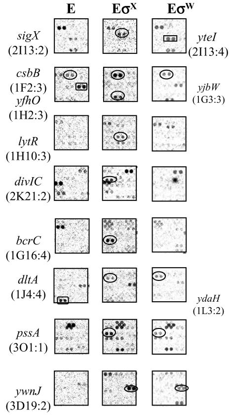
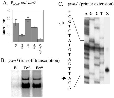
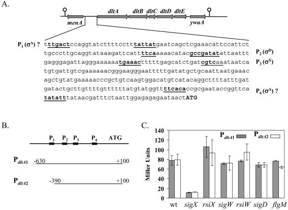
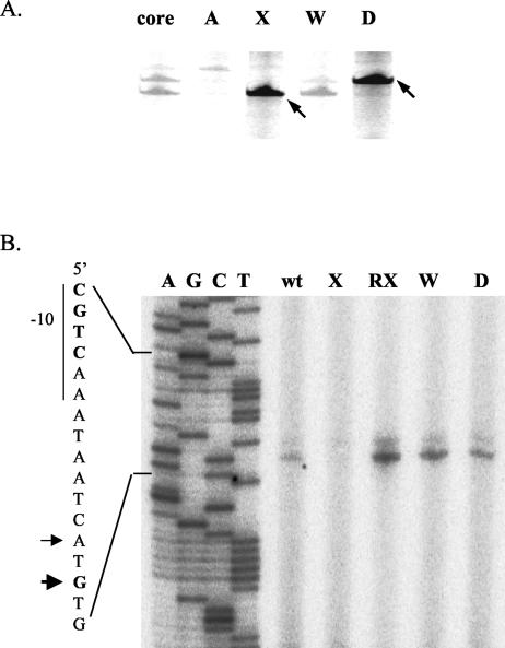
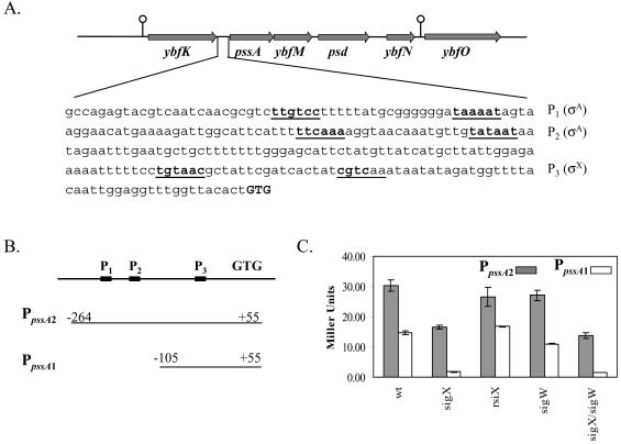
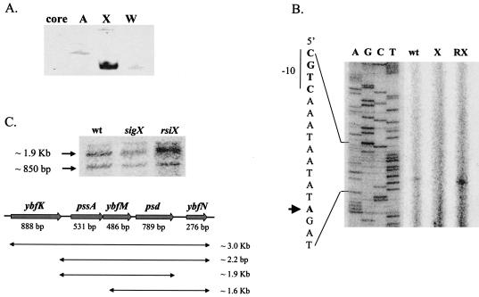
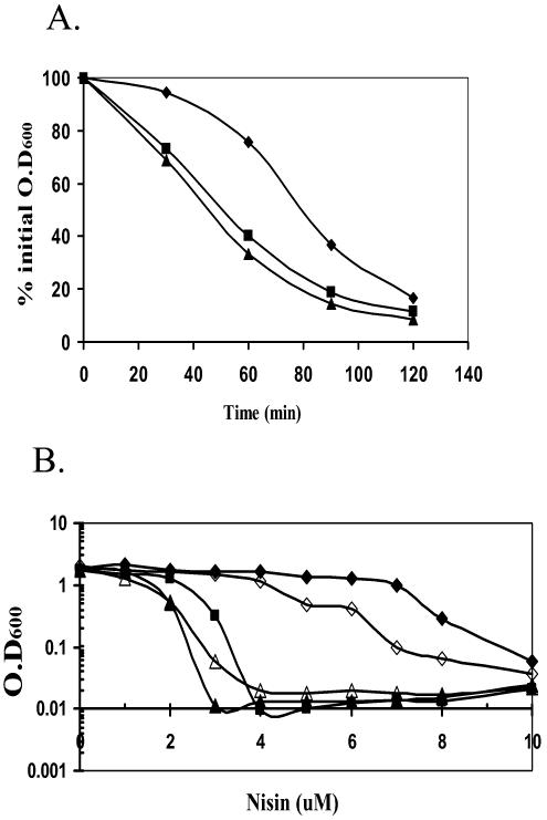
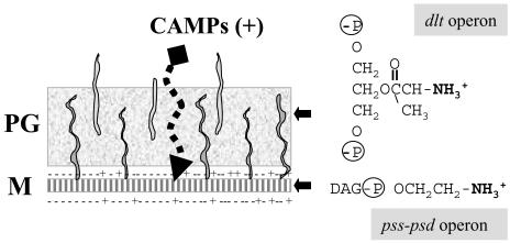
Bacillus subtilis contains seven extracytoplasmic-function sigma factors that activate partially overlapping regulons. We here identify four additional members of the sigma(X) regulon, pbpX (penicillin-binding protein), ywnJ, the dlt operon (D-alanylation of teichoic acids), and the pss ybfM psd operon (phosphatidylethanolamine biosynthesis). Modification of teichoic acids by esterification with D-alanine and incorporation of phosphatidylethanolamine into the cell membrane have a common consequence: in both cases positively charged amino groups are introduced into the cell envelope. The resulting reduction in the net negative charge of the cell envelope has been previously implicated as a resistance mechanism specific for cationic antimicrobial peptides. Consistent with this notion, we find that both sigX and dltA mutants are more sensitive to nisin than wild-type cells. We conclude that activation of the sigma(X) regulon serves to alter cell surface properties to provide protection against antimicrobial peptides.


Figures

FIG.1.
FIG.1.
Identification of σX regulon genes by ROMA. Total B. subtilis chromosomal DNA was digested with EcoRI and transcribed in vitro with core alone (E [left column]) or core with an excess of σX (EσX [central column]). For comparison, the same regions from previous ROMA experiment with EσW are placed in the right column. The σX-regulated genes are apparent in experiments with EσX (ovals). yteI (rectangle) is a σW-dependent gene. Since the core is contaminated with trace amounts of other σ factors, several nonspecific spots appeared on the membrane even in the core-alone experiment. Some spots disappeared or were greatly decreased in abundance upon supplementation with a large molar excess of σX or σW (e.g., yjbW and ydaH, rectangles in left column). Other genes, such as sigX and csbB, which have multiple promoters are found in the RNA population transcribed by core as well as the σX- or σW-supplemented reactions. Three genes identified in this study (dltA, pssA, and ywnJ) are found in both EσX and EσW reactions, but only ywnJ can be transcribed by both σX and σW as confirmed by runoff transcription assays. The location of each gene on the Sigma/GenoSys macroarray is indicated in parentheses.
FIG. 2.
FIG. 2.
Confirmation of the pbpX and ywnJ targets. (A) Expression of PpbpX-cat-lacZ in various genetic backgrounds. Each result is the average for three individual β-galactosidase measurements. (B) In vitro recognition of the putative ywnJ promoter by both the B. subtilis σX (EσX) and the σW (EσW) holoenzymes. The RNAP core enzyme (E) was used as a negative control. (C) RNA generated by runoff transcription using EσX as shown in panel B was used as a template for primer extension mapping of the ywnJ transcription start site (lane X). The same primer was used to sequence this region to index the start site (lanes A, G, C, and T).
FIG. 3.
FIG. 3.
Regulation of the dlt operon by σX. (A) The location of the dltABCDE operon on the B. subtilis chromosome and the dltA promoter region. The σX-dependent promoter (P3), the σD-dependent promoter (P2), and two putative σA-dependent promoters (P1 and P4) are underlined. The translation start codon (ATG) is shown in bold capital letters. (B and C) Graphic presentation of the two PdltA promoter fusions (B) and their activities in various genetic backgrounds (C) (each result is the average and standard deviation from three individual measurements).
FIG. 4.
FIG. 4.
Identification of the σX-dependent promoter for the dlt operon. (A) Runoff transcription from the dltA promoter region in the presence of B. subtilis RNAP core enzyme and the indicated σ factor: σA (A), σX (X), σW (W), or σD (D). In the first lane (core), no σ factor was added in the reaction. Major transcripts are indicated by arrows. (B) Primer extension mapping of the in vivo dltA transcription start site. RNA samples were prepared from wild-type (wt), sigX (X), rsiX (RX), sigW (W), or sigD (D) mutant strains. Equal amounts (100 μg) of total RNA were annealed with radiolabeled oligonucleotide #368 for reverse transcription. The transcription start sites corresponding to the σX-dependent promoter are indicated by arrows.
FIG. 5.
FIG. 5.
Regulation of the pssA ybfM psd operon by σX. (A) Locations of the pssA, ybfM, and psd genes on the B. subtilis chromosome and DNA sequence of the pssA promoter region. The σX-dependent promoter (P3) and two putative σA-dependent promoters (P1 and P2) are underlined. The translation start codon (GTG) is shown in bold capital letters. (B and C) Graphic presentation of the construction of two PpssA promoter fusions (B) and their activities in various genetic backgrounds (C) (each result is the average of three individual measurements).
FIG. 6.
FIG. 6.
Identification of the σX-dependent promoter for the pssA ybfM psd operon. (A) Runoff transcription from the pssA promoter region in the presence of B. subtilis RNAP core enzyme and the indicated σ factor: σA (A), σX (X), or σW (W). In the first lane (core), no σ factor was added in the reaction. (B) Primer extension mapping of the pssA transcription start site. RNA samples were prepared from the wild type (wt) or from sigX (X) or rsiX (RX) mutant strains. Equal amounts (100 μg) of total RNA were annealed with radiolabeled oligonucleotide #408 for reverse transcription. The transcription start site is indicated by the arrow. (C) Northern blot analysis demonstrates that pssA, ybfM, and psd are cotranscribed. The combinations and sizes of possible transcripts are listed. Two bands were observed: the top band is about 1.9 kb, representing the transcript from pssA to psd, while the lower band (∼850 bp) can only be assigned to the psd mRNA, probably due to RNA processing.
FIG. 7.
FIG. 7.
Effects of sigX, dlt, and psd on autolysis and nisin sensitivity. (A) Autolysis rates. B. subtilis CU1065 (wild type; diamonds) and the sigX::spc (squares) and dltA::spc (triangles) mutants were grown to exponential growth phase (OD600, ∼0.7). The cell pellets were washed twice with cold Tris buffer (pH 7.1) and resuspended in 50 mM Tris-HCl buffer (pH 7.1) containing 0.05% Triton X-100. Incubation was at 37°C, and autolysis was monitored by measuring the decrease of OD600 at 30-min intervals. (B) MIC of nisin for the growth of B. subtilis wild-type (closed diamonds), sigX::spc (closed squares), dltA::spc (closed triangles), psd::neo (open diamonds), and dltA psd (open triangle) strains. All strains were grown for 6 h after dilution into LB medium containing the indicated concentration of nisin. This experiment was repeated three times, and representative results are shown.
FIG. 8.
FIG. 8.
Roles of the B. subtilis σX protein in resistance to CAMPs. The B. subtilis cell envelope includes both a cytoplasmic membrane (M) and a thick peptidoglycan layer (PG). Two of the operons controlled by σX are involved in modulating the net charge of the cell envelope. The dlt operon encodes proteins involved in the d-alanylation of both LTA and WTA by esterification of the glycerol moieties with d-alanine. Since both LTA and WTA are glycerol-phosphate copolymers, the introduction of d-alanine esters reduces the net negative charge of the cell wall. Similarly, the cytoplasmic membrane contains an abundance of anionic phospholipids (indicated by −), and the net charge of the membrane can be modulated by the incorporation of neutral constituents, such as glycolipids and the zwitterionic PE. The synthesis of PE requires the products of the pssA ybfM psd operon, which is partially under σX control. The ability of CAMPs to penetrate the cell wall and permeabilize the membrane is reduced by the incorporation of these positively charged groups into the cell envelope.

References

    1. Abachin, E., C. Poyart, E. Pellegrini, E. Milohanic, F. Fiedler, P. Berche, and P. Trieu-Cuot. 2002. Formation of D-alanyl-lipoteichoic acid is required for adhesion and virulence of Listeria monocytogenes. Mol Microbiol. 43:1-14. - PubMed
    1. Akbar, S., and C. W. Price. 1996. Isolation and characterization of csbB, a gene controlled by Bacillus subtilis general stress transcription factor σB. Gene 177:123-128. - PubMed
    1. Boyd, D. A., D. G. Cvitkovitch, A. S. Bleiweis, M. Y. Kiriukhin, D. V. Debabov, F. C. Neuhaus, and I. R. Hamilton. 2000. Defects in D-alanyl-lipoteichoic acid synthesis in Streptococcus mutans results in acid sensitivity. J. Bacteriol. 182:6055-6065. - PMC - PubMed
    1. Brutsche, S., and V. Braun. 1997. SigX of Bacillus subtilis replaces the ECF sigma factor FecI of Escherichia coli and is inhibited by RsiX. Mol. Gen. Genet. 256:416-425. - PubMed
    1. Bsat, N., A. Herbig, L. Casillas-Martinez, P. Setlow, and J. D. Helmann. 1998. Bacillus subtilis contains multiple Fur homologues: identification of the iron uptake (Fur) and peroxide regulon (PerR) repressors. Mol. Microbiol. 29:189-198. - PubMed

Publication types

MeSH terms

LinkOut - more resources