. 2004 Feb 4;24(5):1070-8.
doi: 10.1523/JNEUROSCI.4719-03.2004.

Ephrin-A5 exerts positive or inhibitory effects on distinct subsets of EphA4-positive motor neurons

Affiliations

Ephrin-A5 exerts positive or inhibitory effects on distinct subsets of EphA4-positive motor neurons

Johann Eberhart et al. J Neurosci. .

Abstract

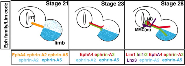
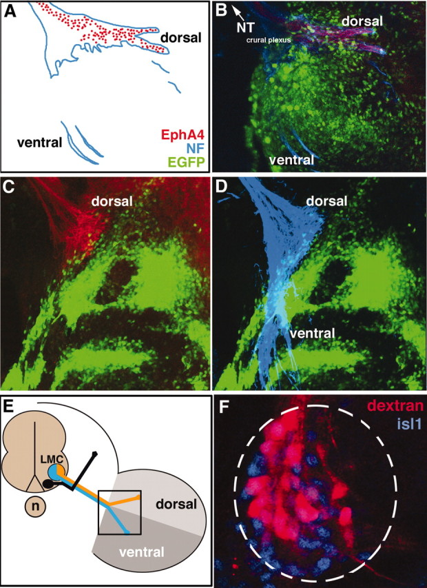
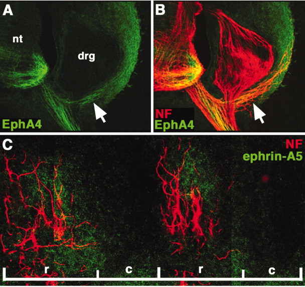
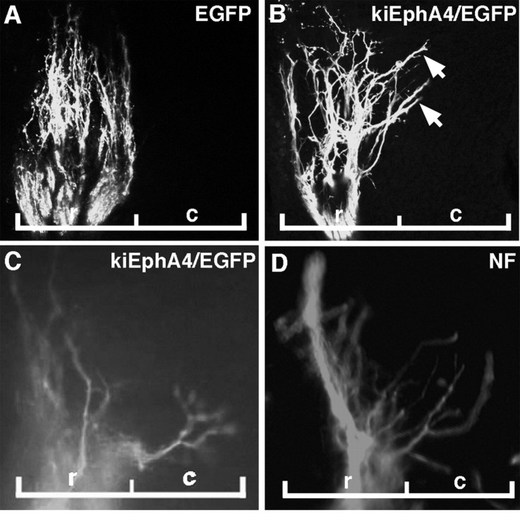
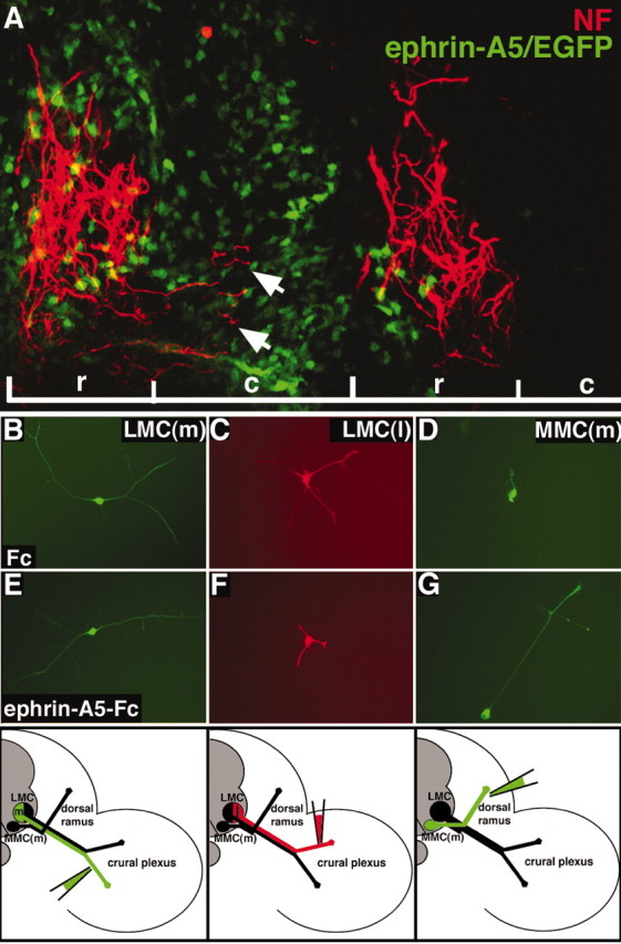
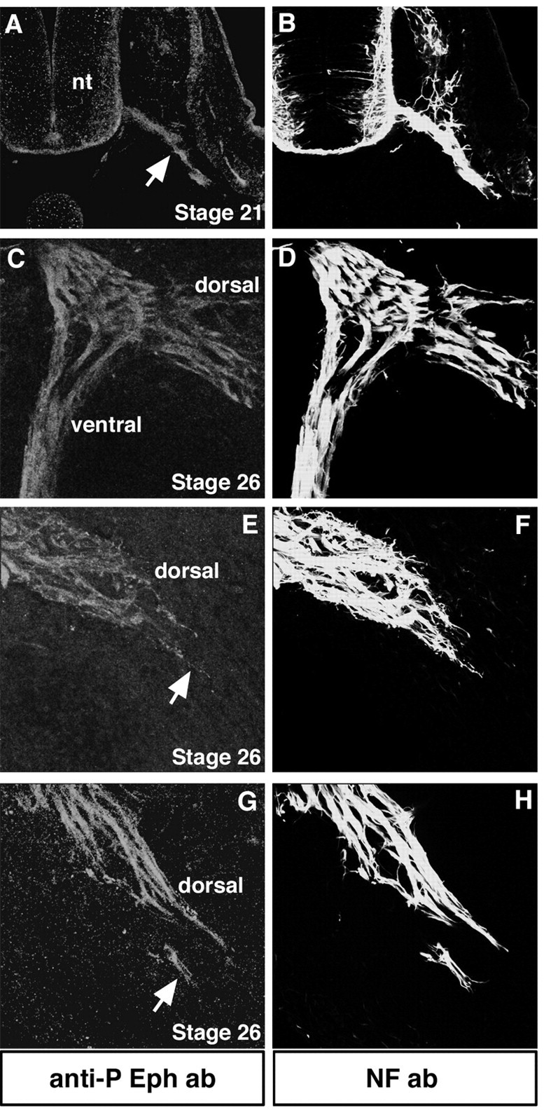
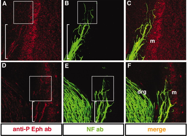
Eph receptor tyrosine kinases and ephrins are required for axon patterning and plasticity in the developing nervous system. Typically, Eph-ephrin interactions promote inhibitory events; for example, prohibiting the entry of neural cells into certain embryonic territories. Here, we show that distinct subsets of motor neurons that express EphA4 respond differently to ephrin-A5. EphA4-positive LMC(l) axons avoid entering ephrin-A5-positive hindlimb mesoderm. In contrast, EphA4-positive MMC(m) axons extend through ephrin-A5-positive rostral half-sclerotome. Blocking EphA4 activation in MMC(m) neurons or expanding the domain of ephrin-A5 expression in the somite results in the aberrant growth of MMC(m) axons into the caudal half-sclerotome. Moreover, premature expression of EphA4 in MMC(m) neurons leads to a portion of their axons growing into novel ephrin-A5-positive territories. Together, these results indicate that EphA4-ephrin-A5 signaling acts in a positive manner to constrain MMC(m) axons to the rostral half-sclerotome. Furthermore, we show that Eph activation localizes to distinct subcellular compartments of LMC(l) and MMC(m) neurons, consistent with distinct EphA4 signaling cascades in these neuronal subpopulations.


Figures

Figure 1.
Figure 1.
The expression of Eph family members and the Lim code during the development of motor axon projections to target muscle. All diagrams illustrate expression patterns at the level of the crural plexus in the hindlimb. Left, All motor axons express EphA4, ephrin-A2, and ephrin-A5 (gold) when they reach the base of the hindlimb at stage 21. Ephrin-A2 is diffusely expressed across the hindlimb mesoderm, whereas ephrin-A5 is restricted to the ventral hindlimb. Middle, As motor axons initiate sorting in the plexus at stage 23, EphA4 is segregated to the forming dorsal nerve trunk (red) and presumably downregulated on the forming ventral nerve trunk. Ephrin-A5 protein is absent on motor axons whereas ephrin-A2 remains on all motor axons (red, green). Right, When the adult pattern of axon projections have formed, motor neurons in the LMC(l) (green) express Lim1, EphA4, and ephrin-A2 and enter the dorsal hindlimb, which lacks ephrins. Motor neurons in the LMC(m) (red) project to the ventral hindlimb, which is rich in ephrins (blue) and express Isl1/2 and ephrin-A2, but not EphA4. MMC(m) neurons (purple) project to epaxial muscle and express Lhx3, Isl1/2, and EphA4 (this study).
Figure 5.
Figure 5.
MMC(m) axons extend inappropriately into novel, ephrin-A5-positive territories when EphA4/EGFP is prematurely expressed. A, Transverse section through the neural tube (nt) 2 d after electroporation of EphA4/EGFP construct (green) into MMC(m) neurons shows that a single Isl1/2-positive axon (arrow) misprojects into the dorsal neural tube. B, Transverse section, labeled with ephrin-A5 (green) and isl1/2 (red) antibodies, shows that ephrin-A5 is strongly expressed in the dorsal half (*) neural tube (nt), lateral to recently postmitotic neurons in the ventral neural tube (arrowhead), and in the forming DRG (arrow) at the stage when MMC(m) axons misproject into the neural tube (A) or DRG (C,D). C, D, Transverse section showing that an EphA4/EGFP-positive axon (arrow) is found in the dorsal root ganglia (drg). Red, NF antibody staining; green, EGFP signal. E, F, High magnification of transverse section through ventral neural tube showing that a MMC(m) neuron cell body (arrow) has been backfilled from an aberrant axon in the DRG. Note that the LMC is devoid of dextran-labeled neurons. Green, EGFP signal; red, dextran amines; blue, Lhx3 antibody staining to label MMC(m) neurons.
Figure 2.
Figure 2.
EphA4+ LMC(l) axons are prohibited from limb entry by ectopic expression of ephrin-A5 in limb mesoderm. A, B, Schematic diagram and image showing that EphA4+ dorsal axons (red) and neurofilament antibody-labeled axons (blue) of the crural plexus enter the limb normally when EGFP (green) is expressed in limb mesoderm, controls. C, When ephrin-A5/EGFP is ectopically expressed in limb mesoderm, dorsal EphA4+ axons do not enter the limb. D, Neurofilament (NF) antibody reveals that axons in the ventral nerve trunk enter normally in the presence of ectopic ephrin-A5/EGFP. E, Schematic diagram showing the views of LMC axons depicted in A–D, in boxed area. F, Backfills of the ventral nerve trunk in embryos where ephrin-A5 was ectopically expressed in limb mesoderm reveals that LMC(m) cell bodies are labeled, indicating that axons in the ventral nerve trunk have sorted properly.
Figure 3.
Figure 3.
EphA4 localizes predominantly to MMC(m) axon shafts; ephrin-A5 is expressed in rostral half-sclerotome. A, B, Transverse section showing that EphA4 protein (green) strongly marks MMC(m) axon shafts (arrow) that lie lateral to the dorsal root ganglia (drg), at stage 26. Neurofilament (NF) antibody staining labels all axons (red). C, Sagittal section showing that ephrin-A5 protein (green) localizes primarily to the rostral half-sclerotome, occupied by MMC(m) axons that are labeled by NF antibody (red). nt, Neural tube; r, rostral half-sclerotome; c, caudal half-sclerotome.
Figure 4.
Figure 4.
MMC(m) axons grow aberrantly into the caudal half-sclerotome when EphA4 signaling is blocked. All images are sagittal sections through MMC(m) axons, at the level of a single somite. A, At stage 26, MMC(m) axons grow normally in the rostral half-sclerotome when EGFP is expressed in MMC(m) neurons, controls. B, At stage 26, some MMC(m) axons extend abnormally (arrows) into the caudal half-sclerotome when kinase-inactive EphA4/EGFP is expressed in MMC(m) neurons to block EphA4 phosphorylation. C, D, At stage 28, MMC(m) axons are localized inappropriately in the caudal half-sclerotome when EphA4 signaling is blocked. Green, EGFP signal; red, NF antibody labeling. r, rostral half-sclerotome, c, caudal half-sclerotome.
Figure 6.
Figure 6.
Neuron responses to ephrin-A5 are distinct. A, Sagittal section showing that NF+ MMC(m) axons (red) grow aberrantly (arrows) into the caudal half-sclerotome when the domain of ephrin-A5 expression (green) in the sclerotome is expanded. r, Rostral half-sclerotome; c, caudal half-sclerotome. B, E, LMC(m) neurons that are EphA4-demonstrate robust growth on ephrin-A5 or in controls (Fc). C, F, The growth of LMC(l) neurons that express EphA4 is reduced on ephrin-A5 substrates, compared with controls (C). D, G, MMC(m) neurons display poor growth in control cultures but exuberant neurite growth on ephrin-A5 substrates. Bottom, Schematic diagrams showing that retrograde labeling of the ventral nerve trunk to label LMC(m) neurons, of the dorsal nerve trunk to mark LMC(l) neurons, and of the dorsal ramus to label MMC(m) neurons is precise.
Figure 7.
Figure 7.
Eph phosphorylation localizes to the axon shafts and distal tips of LMC neurons in vivo. All transverse sections were labeled with anti-phosphorylated Eph antibody to detect Eph activation (red) and NF antibody to label all axons (green). A, B, At stage 21, before motor axons arrive to the base of the limb, they exhibit Eph phosphorylation (arrow). nt, Neural tube. C, D, At the level of the crural plexus at stage 26, dorsal and ventral projecting axons display Eph phosphorylation. E–H, Distal tips of LMC(l) axons (arrows) show Eph phosphorylation.
Figure 8.
Figure 8.
MMC(m) neurons show Eph phosphorylation at their axon shafts but negligible signal at their distal axon tips in vivo. High-magnification views of transverse sections through MMC(m) axons labeled with anti-phosphorylated Eph antibody (red) and NF antibody (green).A–C, MMC(m) axon shafts (bracket inA) exhibit Eph activation, whereas distal tips show extremely weak Eph activation (boxed area). D–F, In another embryo, MMC(m) axon shafts show strong activation of Eph (bracket in A) but very weak signal localized to their distal tips (boxed area). drg, Dorsal root ganglia; m, myotome.

References

    1. Brittis PA, Qiang L, Flanagan JG (2002) Axonal protein synthesis provides a mechanism for localized regulation at an intermediate target. Cell 110: 223–235. - PubMed
    1. Campbell DS, Holt CE (2001) Chemotropic responses of retinal growth cones mediated by rapid local protein synthesis and degradation. Neuron 32: 1013–1026. - PubMed
    1. Dickson BJ (2002) Molecular mechanisms of axon guidance. Science 298: 1959–1964. - PubMed
    1. Eberhart J, Swartz M, Koblar SA, Pasquale EB, Tanaka H, Krull CE (2000) Expression of EphA4, ephrin-A2 and ephrin-A5 during axon outgrowth to the hindlimb indicates potential roles in pathfinding. Dev Neurosci 22: 237–250. - PubMed
    1. Eberhart J, Swartz ME, Koblar SA, Pasquale EB, Krull CE (2002) EphA4 constitutes a population-specific guidance cue for motor neurons. Dev Biol 247: 89–101. - PubMed

Publication types

MeSH terms