. 2004 Feb 17;101(7):2100-5.
doi: 10.1073/pnas.0306448101. Epub 2004 Feb 4.

A capsid protein of nonenveloped Bluetongue virus exhibits membrane fusion activity

Affiliations

A capsid protein of nonenveloped Bluetongue virus exhibits membrane fusion activity

Mario Forzan et al. Proc Natl Acad Sci U S A. .

Abstract

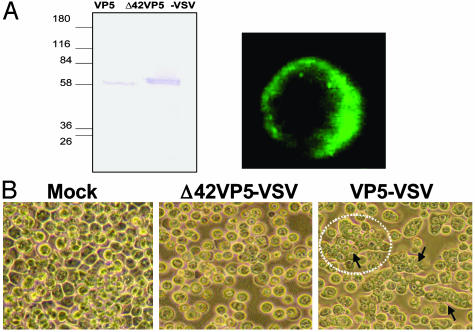
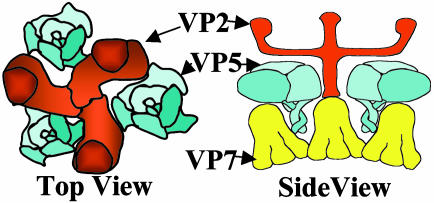
The outer capsid layer of Bluetongue virus, a member of the nonenveloped Reoviridae family, is composed of two proteins, a receptor-binding protein, VP2, and a second protein, VP5, which shares structural features with class I fusion proteins of enveloped viruses. In the replication cycle of Bluetongue virus VP5 acts as a membrane permeabilization protein that mediates release of viral particles from endosomal compartments into the cytoplasm. Here, we show that VP5 can also act as a fusion protein and induce syncytium formation when it is fused to a transmembrane anchor and expressed on the cell surface. Fusion activity is strictly pH-dependent and is triggered by short exposure to low pH. No cell-cell fusion is observed at neutral pH. Deletion of the first 40 amino acids, which can fold into two amphipathic helices, abolishes fusion activity. Syncytium formation by VP5 is inhibited in the presence of VP2 when it is expressed in a membrane-anchored form. The data indicate an interaction between the outer capsid protein VP2 and VP5 and show that VP5 undergoes pH-dependent conformational changes that render it capable of interacting with cellular membranes. More importantly, our data show that a membrane permeabilization protein of a nonenveloped virus can evolve into a fusion protein by the addition of an appropriate transmembrane anchor. The results strongly suggest that the mechanism of membrane permeabilization by VP5 and membrane fusion by viral fusion proteins require similar structural features and conformational changes.


Figures

Fig. 1.
Fig. 1.
Construction and expression of membrane-anchored VP2 and VP5. (A) Baculovirus transfer vectors were constructed in which the coding sequences of VP2 and VP5 of BTV10 were fused in-frame to the signal peptide of the baculovirus gp64 and the C-terminal part of VSV G. (B) SDS/10% PAGE of insect cells infected with AcVP2-VSV (Left) and AcVP5-VSV (Right). Lanes: 1, cell lysate of uninfected Sf9 cells; 2, purified VP2 or VP5; 3, lysate of insect cells infected for 42 h with AcVP2-VSV or AcVP5-VSV. (C) Flow cytometric analysis. Sf9 cells were infected with AcVP2-VSV and AcVP5-VSV, were stained with polyclonal antisera and FITC-conjugated secondary antibodies, and were analyzed on a FACScan flow cytometer. Mock-infected cells were stained with the same antibodies as a control for nonspecific binding. (D) Immunofluorescence assay. Sf9 cells were coinfected at an moi of 2.5 for 42 h. The cells were then labeled under nonpermeabilizing conditions with polyclonal antisera against VP2 and VP5, followed by FITC-conjugated secondary antibodies, and were visualized on a Nikon fluorescence microscope. (E) Confocal microscopy. Sf9 cells were infected and stained as described above, except that tetramethylrhodamine isothiocyanate-conjugated secondary antibodies were used to label VP2. Pictures were taken on a Zeiss LSM510 microscope.
Fig. 2.
Fig. 2.
Cell viability assay. (A) Sf9 cells were infected at an moi of 5 for 48 h and were then incubated in medium containing 0.2% Trypan blue. Pictures were taken on a Nikon light microscope to visualize dead cells. (Aa) Uninfected cells. (Ab) Cells expressing cytoplasmic VP2. (Ac) Cells expressing cytoplasmic VP5. (Ad) Cells expressing membrane-anchored VP2. (Ae) Cells expressing membrane-anchored VP5. (Af) Cells expressing both VP2-VSV and VP5-VSV. (B) The experiment described in A was repeated and the number of dead cells was counted 24, 48, and 60 h after infection. The percentage of dead cells is shown in the graph.
Fig. 3.
Fig. 3.
Fusogenic activity of VP5-VSV. (A) Sf9 cells were infected for 48 h at an moi of 2.5 and were then exposed to pH 5.0 for 2 min, after which the low-pH buffer was replaced by normal growth medium. Pictures were taken on an inverted light microscope at different time points after the pH shift. Uninfected cells (Left) and infected cells after 4 h (Center) and 7 h (Right) pH shift are shown. (B) Insect cells were infected for 24, 48, and 60 h, after which they were exposed to pH 5.0 as described above. Pictures were taken before and 4 h after the pH shift, and the number of cells per syncytium were counted at each time point (C).
Fig. 4.
Fig. 4.
Membrane-anchored VP2 lacks fusogenic activity and inhibits VP5 activity. (A) Sf9 cells were infected with AcVP2-VSV at an moi of 2.5. The infected cells were exposed to pH 5.0 at 24, 48, and 60 h after infection, and pictures were taken before and 4 h after the pH shift as described above. (B) Insect cells were coinfected with AcVP2-VSV and AcVP5-VSV each at an moi of 2.5. The fusogenic activity was checked at 48 h after infection as described above, and pictures were taken before and 4 h after the pH shift.
Fig. 5.
Fig. 5.
Expression and fusion activity of membrane-anchored VP5 lacking the amphipathic helices. A recombinant baculovirus AcΔ42VP5-VSV was generated expressing a membrane-anchored mutant of VP5 in which the first 42 amino acids were deleted. (A) Sf9 cells were infected with the recombinant virus and protein expression was monitored by SDS/PAGE 24 h after infection (Left). Confocal microscopy confirmed membrane display on infected Sf9 cells (Right). (B) Insect cells were infected with AcΔ42VP5-VSV for 48 h, and syncytium formation was monitored 4 h after exposure to pH 5. Note that AcΔ42VP5-VSV exhibited no detectable fusion, in comparison with full-length AcVP5-VSV.
Fig. 6.
Fig. 6.
Schematic diagrams of a section of a BTV particle. The schematics show the organizations of the three trimeric major proteins, VP2, VP5, and VP7, depicted from image reconstruction of cryo-electron microscopy analysis and the arrangement of VP5 in relation to VP2 (top view) or VP2 and VP7 (side view). Note that VP2 shrouds VP5 under normal physiological conditions.

References

    1. Cohen, F. S. & Melikyan, G. B. (2001) Nat. Struct. Biol. 8, 653-655. - PubMed
    1. Peisajovich, S. G. & Shai, Y. (2002) Trends Biochem. Sci. 27, 183-190. - PubMed
    1. Tamm, L. K., Han, X., Li, Y. & Lai, A. L. (2002) Biopolymers 66, 249-260. - PubMed
    1. Jahn, R., Lang, T. & Sudhof, T. C. (2003) Cell 112, 519-533. - PubMed
    1. Weissenhorn, W., Dessen, A., Calder, L. J., Harrison, S. C., Skehel, J. J. & Wiley, D. C. (1999) Mol. Membr. Biol. 16, 3-9. - PubMed

Publication types

LinkOut - more resources