. 2004 Apr 13;101(15):5318-24.
doi: 10.1073/pnas.0307824100. Epub 2004 Feb 4.

Small potassium ion channel proteins encoded by chlorella viruses

Affiliations

Small potassium ion channel proteins encoded by chlorella viruses

Ming Kang et al. Proc Natl Acad Sci U S A. .

Abstract

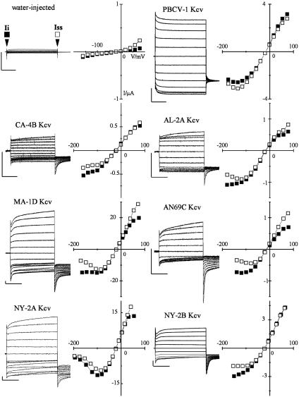
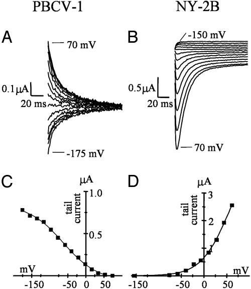
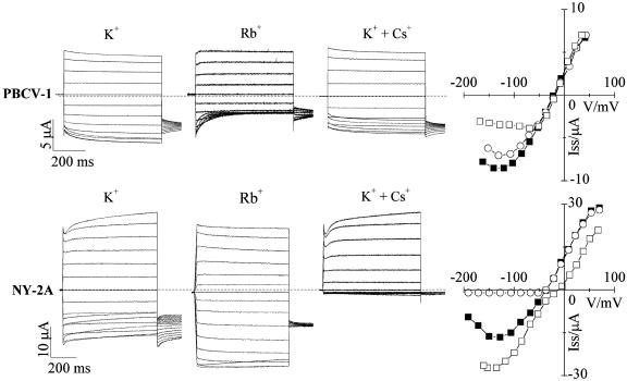
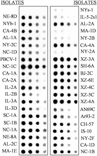
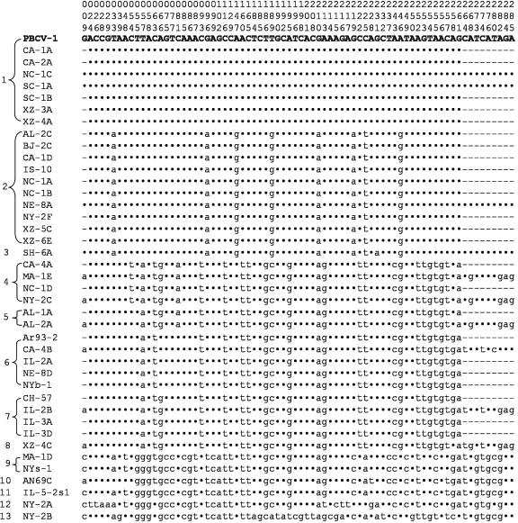
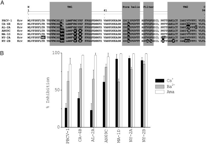
Kcv, a 94-aa protein encoded by Paramecium bursaria chlorella virus 1, is the smallest known protein to form a functional potassium ion channel and basically corresponds to the "pore module" of potassium channels. Both viral replication and channel activity are inhibited by the ion channel blockers barium and amantadine but not by cesium. Genes encoding Kcv-like proteins were isolated from 40 additional chlorella viruses. Differences in 16 of the 94 amino acids were detected, producing six Kcv-like proteins with amino acid substitutions occurring in most of the functional domains of the protein (N terminus, transmembrane 1, pore helix, selectivity filter, and transmembrane 2). The six proteins form functional potassium selective channels in Xenopus oocytes with different properties including altered current kinetics and inhibition by cesium. The amino acid changes together with the different properties observed in the six Kcv-like channels will be used to guide site-directed mutations, either singularly or in combination, to identify key amino acids that confer specific properties to Kcv.


Figures

Fig. 4.
Fig. 4.
All Kcv variants form functional K+ channels in Xenopus oocytes. Expression of PBCV-1 Kcv and the six Kcv-like channels in Xenopus oocytes. Currents were recorded in 50 mM KCl from a representative oocyte injected either with water or with the cRNA of the indicated protein. Standard voltage protocol was: holding potential, –20 mV; steps of –20 mV from +80 mV to ≤–160 mV; postpotential, –80 mV. The corresponding I–V relation (Right) shows the instantaneous (Ii, ▪) and the steady-state (Iss, □) currents, sampled at the beginning and end of the voltage pulse, as indicated by arrows in the water-injected sample. Vertical bar, 1 μA; horizontal bar, 200 ms.
Fig. 5.
Fig. 5.
Currents conducted by Kcv variants exhibit different kinetics. Shown is tail current analysis of PBCV-1 Kcv (A) and NY-2B Kcv (B), enlargement of the tail currents of PBCV-1 Kcv and NY-2B Kcv from Fig. 4. Tail currents were collected at –80 mV after stepping to test voltages between the indicated values. (C and D) Activation curves obtained from the tail current values (after subtraction of the time-independent current component) plotted against the conditioning voltages. Data were fitted with a Boltzmann-derived equation, Y = A1–A2/(1 + exp ((x-x0)/dx))) + A2. The value for half-maximum activation, x0, is –62.5 mV for PBCV-1 Kcv (C) and 53.2 mV for NY-2B (D). The inverse slope factor, dx, is 39.2 mV for PBCV-1 Kcv and –35 mV for NY-2B.
Fig. 6.
Fig. 6.
Kcv variants show different cation dependencies. Shown is a comparison of current properties of PBCV-1 Kcv (Upper) and NY-2A Kcv (Lower) in the presence of different cations. First column, reference currents recorded in K+. Second column, opposite effects of Rb+ on the kinetics and the inward current. Third column, different degrees of voltage-dependent inhibition of K+ current by Cs+. Fourth column, I–V plots reporting steady-state currents (▪) 50 mM K+, (□) 50 mM Rb+, (○)50mMK+ + 10 mM Cs+. Standard voltage protocol is as in Fig. 4.
Fig. 1.
Fig. 1.
The kcv-like gene is ubiquitous in chlorella viruses. Hybridization of the PBCV-1 kcv gene to DNA isolated from 40 viruses that infect Chlorella NC64A. The spots contain 1.0, 0.5, 0.25, and 0.125 μg of DNA (left to right).
Fig. 2.
Fig. 2.
Diversity of kcv gene among chlorella viruses. Differences in the 285 nucleotide coding regions (including stop codons) of the kcv gene from 40 chlorella viruses as compared with the PBCV-1 kcv gene are shown. Only the sites that had a nucleotide change, numbered at the top, are listed. A dot indicates no change, and a dash means not determined.
Fig. 3.
Fig. 3.
Diversity of Kcv proteins and sensitivity of corresponding viruses to channel inhibitors. (A) Alignment of the seven different Kcv proteins. Each protein is named after a virus representative from each group. Amino acid substitutions, compared to PBCV-1 Kcv, are highlighted in black. The assignment of putative structure domains is based on an alignment with PBCV-1 Kcv and KcsA (5). TM1 and TM2, outer and inner transmembrane domains, respectively. (B) Plaque inhibition of chlorella viruses by potassium channel inhibitors amantadine, barium, and cesium.

Similar articles

Cited by

References

    1. Van Etten, J. L. & Meints, R. H. (1999) Annu. Rev. Microbiol. 53, 447–494. - PubMed
    1. Van Etten, J. L. (2003) Annu. Rev. Genet. 37, 153–195. - PubMed
    1. Plugge, B., Gazzarrini, S., Nelson, M., Cerana, R., Van Etten, J. L., Derst, C., DiFrancesco, D., Moroni, A. & Thiel, G. (2000) Science 287, 1641–1644. - PubMed
    1. Moroni, A., Viscomi, C., Sangiorgio, V., Pagliuca, C., Meckel, T., Horvath, F., Gazzarrini, S., Valbuzzi, P., Van Etten, J. L., DiFrancesco, D. & Thiel, G. (2002) FEBS Lett. 530, 65–69. - PubMed
    1. Gazzarrini, S., Severino, M., Lombardi, M., Morandi, M., DiFrancesco, D., Van Etten, J., Thiel, G. & Moroni, A. (2003) FEBS Lett. 552, 12–16. - PubMed

Publication types

LinkOut - more resources