Review
. 2004 Apr;24(4):650-7.
doi: 10.1161/01.ATV.0000120376.09047.fe. Epub 2004 Feb 5.

Expansive arterial remodeling: location, location, location

Affiliations
Review

Expansive arterial remodeling: location, location, location

Gerard Pasterkamp et al. Arterioscler Thromb Vasc Biol. 2004 Apr.

Abstract

The artery is a dynamic organ capable of changing its geometry in response to atherosclerotic plaque formation. Expansion of the vessel diameter retards luminal narrowing and is considered a compensatory response. However, the expansive remodeling response is a "wolf in sheep's clothes," because expansion is associated with the presence of inflammatory cells, proteolysis, and a thrombotic plaque phenotype. The prevalence and clinical presentation of expansively remodeled lesions may differ among vascular beds. However, it is evident that all types of atherosclerotic arterial expansive lesions share the presence of inflammatory cells and subsequent protease activities. The potential role of inflammation and protease activity in the development of the different remodeling modes is discussed.


Figures

Figure 1
Figure 1
Schematic presentation of the different remodeling modes. The mode and extent of arterial remodeling is mostly based on a comparison with 1 or 2 reference sites. In postmortem studies, often the vessel area in the section with the least amount of plaque (R1) is used as a reference. In intravascular ultrasound studies, the average of the vessel areas of a proximal (R1) and distal site (R) with an angiographic normal lumen is used as a reference. Expansive remodeling (E) is then found present when: (vessel area R1+R2)/2> vessel area culprit lesion. However, constrictive remodeling [C] is evident when: (vessel area R1+R2)/2< vessel area culprit lesion. Expansive remodeling is often associated with the presence of atheroma and inflammatory cells (see text).
Figure 2
Figure 2
Distribution of delta plaque area (PA), delta vessel area (VA), and delta lumen (LA) are in 609 coronary lesions before intervention. Delta was defined as difference between measured values of the lesion site and the average value of both reference sites as visualized with intravascular ultrasound. The reference site appeared normal on the angiogram and no major side branches originated between the reference sites and the lesion sites. We studied the distribution of the axial changes in plaque area and vessel area and their relative contributions to changes of luminal area for the 609 culprit coronary lesions. Both axial changes in plaque area and vessel area were normally distributed. A comparable width of the 95% confidence intervals was observed for segmental variation in plaque area and vessel area (−1.02 mm2, +11.56 mm2) and (−6.44 mm2, +5.04 mm2), respectively. (Figure obtained with permission from Pasterkamp G, de Kleijn D, Fitzgerald P. Expansive remodeling, a sheep in wolf’s clothes. J Vasc Res. 2002;39:514–523).
Figure 3
Figure 3
Quartiles of different degrees of remodeling against lumen, vessel, plaque, and media area. A total of 1595 sections were obtained from 112 femoral arteries at regular intervals of 0.5 cm. In each artery, the cross-section with the least amount of plaque was considered the reference. A percent increase in vessel area was calculated for other sections compared with this reference. Cross-sections were divided in the quartiles based on this relative remodeling percent. The lumen, vessel, plaque, and media of the reference sites were 21.6 ±9.1, 33.1±15.4, 13.1±9.4, and 7.0±3.4 mm2, respectively. *P<0.05.
Figure 4
Figure 4
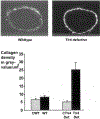
Collagen density within the wall of contralateral (left) mouse carotid arteries before and after ligation of the right carotid artery. Top panels are digital gray value images of a picro Sirius red image with circularly polarized light. Left is wild-type left carotid artery 28 days after ligation. Right is Tlr4-deficient left carotid artery 28 days after ligation. Bottom graph shows quantification of collagen density in left carotid In gray bars, left carotid arteries before ligation in wild-type (cWT) and Tlr4-deficient mice (cTlr4 def) are shown. In black bars, left carotid arteries 28 days after right carotid ligation in wild-type (WT) and Tlr4-deficient mice (Tlr4 def) are shown. N=14 to 17 mice per group, *P<0.05

References

    1. Langille BL, O’Donnell F. Reductions in arterial diameter produced by chronic decreases in blood flow are endothelium-dependent. Science 1986;231:405–407. - PubMed
    1. Glagov S, Weisenberg E, Zarins CK, Stankunavicius R, Kolettis G. Compensatory enlargement of human atherosclerotic coronary arteries. N Engl J Med 1987;316:1371–1375. - PubMed
    1. Post MJ, de Smet BJ, van der Helm Y, Borst C, Kuntz RE. Arterial remodeling after balloon angioplasty or stenting in an atherosclerotic experimental model. Circulation 1997;96:996–1003. - PubMed
    1. Pasterkamp G, Wensing PJW, Post MJ, Hillen B, Mali WPTM, Borst C. Paradoxical arterial wall shrinkage contributes to luminal narrowing of human atherosclerotic femoral arteries. Circulation 1995;91:1444–1449. - PubMed
    1. Nishioka T, Luo H, Eigler NL, Berglund H, Kim C-J, Siegel RJ. Contribution of inadequate compensatory enlargement to development of human coronary artery stenosis: an in vivo intravascular ultrasound study. J Am Coll Cardiol 1996;27:1571–1576. - PubMed

Publication types

MeSH terms