. 2004 May;18(5):1158-70.
doi: 10.1210/me.2003-0442. Epub 2004 Feb 5.

Activin regulation of the follicle-stimulating hormone beta-subunit gene involves Smads and the TALE homeodomain proteins Pbx1 and Prep1

Affiliations

Activin regulation of the follicle-stimulating hormone beta-subunit gene involves Smads and the TALE homeodomain proteins Pbx1 and Prep1

Janice S Bailey et al. Mol Endocrinol. 2004 May.

Abstract

FSH is critical for normal reproductive function in both males and females. Activin, a member of the TGFbeta family of growth factors, is an important regulator of FSH expression, but little is known about the molecular mechanisms through which it acts. We used transient transfections into the immortalized gonadotrope cell line LbetaT2 to identify three regions (at -973/-962, -167, and -134) of the ovine FSH beta-subunit gene that are required for full activin response. All three regions contain homology to consensus binding sites for Smad proteins, the intracellular mediators of TGFbeta family signaling. Mutation of the distal site reduces activin responsiveness, whereas mutation of either proximal site profoundly disrupts activin regulation of the FSHbeta gene. These sites specifically bind LbetaT2 nuclear proteins in EMSAs, and the -973/-962 site binds Smad4 protein. Interestingly, the protein complex binding to the -134 site contains Smad4 in association with the homeodomain proteins Pbx1 and Prep1. Using glutathione S-transferase interaction assays, we demonstrate that Pbx1 and Prep1 interact with Smads 2 and 3 as well. The two proximal activin response elements are well conserved across species, and Pbx1 and Prep1 proteins bind to the mouse gene in vivo. Furthermore, mutation of either proximal site abrogates activin responsiveness of a mouse FSHbeta reporter gene as well, confirming their functional conservation. Our studies provide a basis for understanding activin regulation of FSHbeta gene expression and identify Pbx1 and Prep1 as Smad partners and novel mediators of activin action.


Figures

Fig. 1
Fig. 1
Full Activin Responsiveness of the FSHβ Gene Requires Regions between −985 and −751, and between −401 and −108 Transient transfections were performed in LβT2 cells with reporter plasmids containing the indicated gene regions controlling luciferase expression. A Rous sarcoma virus-βGal plasmid was cotransfected, and luciferase activity was normalized to β-galactosidase activity to control for transfection efficiency. For each reporter, relative light unit (RLU) values for activin-treated samples were divided by vehicle-treated controls to yield fold activation. Data are the means ± SEM of three independent experiments, each performed in triplicate. *, Activin-treated values that differ significantly from vehicle-treated controls; #, fold activation that differs significantly from that of the previous truncation. A, Activin responsiveness of reporter plasmids containing either the full-length −4741/+759 region of the oFSHβ gene or a 5′-deletion to −985 was assayed. B, Activin responsiveness of reporter plasmids containing either the −985/+759 region or the indicated 5′-deletions was assayed. To control for differences in activin stimulation between different batches of LβT2 cells, fold activation for each reporter was normalized to that of the −985 oFSHβ-Luc plasmid, which was set at 1.
Fig. 2
Fig. 2
Mutation of Putative SBEs Disrupts Activin Responsiveness of the FSHβ Gene A, Sites in the −985 regulatory region with homology to the SBE consensus sequence. Nucleotides in bold indicate bases mutated for use in subsequent transfection and EMSA experiments (base changes are shown in lowercase letters in sequences listed in Table 1). B, Activin responsiveness of reporter plasmids containing either the wild-type −985 region of the oFSHβ gene or the indicated point mutations was assayed in transiently transfected LβT2 cells. Data are the means ± SEM of three independent experiments, each performed in triplicate, and were normalized as described in Fig. 1. *, Values that differ significantly from vehicle-treated control; #, fold activation with activin treatment that differs significantly from fold activation of the wild-type −985 reporter. RLU, Relative light units.
Fig. 3
Fig. 3
Smad4 Binds to the −973/−962 and −453/−442 Regions of the FSHβ Gene EMSA was performed using LβT2 nuclear extract and wild-type (WT) or mutant −973/−962 oligonucleotide probes (panel A) or wild-type or mutant −453/−442 oligonucleotide probes (panel B). Wild-type or mutant competitor oligonucleotides and antibodies (Ab) were included in the reactions as indicated. Specific protein complexes are indicated by arrows, and shifted complexes induced by inclusion of an antibody to Smad4 are indicated by arrowheads. Detection of the specific complexes required a week-long autoradiographic exposure, which resulted in overexposure of the free probe bands. To enable comparison of the specific activity of the individual probes, the free probe panels are from an overnight exposure of the same gels.
Fig. 4
Fig. 4
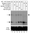
Specific LβT2 Nuclear Proteins Bind to the −167 Region of the FSHβ Gene EMSA was performed using LβT2 nuclear extract and either the wild-type (WT) or mutant −167 oligonucleotide probe. Wild-type or mutant competitor oligonucleotides and antibodies (Ab) were included in the reactions as indicated. Specific protein complexes are indicated by arrows, and the complex disrupted by the mutation is indicated by an arrowhead.
Fig. 5
Fig. 5
Pbx1 and Prep1 Bind to the −134 Region of the FSHβ Gene EMSA was performed using LβT2 nuclear extract and either the wild-type (WT) or mutant −134 oligonucleotide probe. A, Wild-type or mutant competitor oligonucleotides and antibodies (Ab) were included in the reactions as indicated. Pbx1a/Prep1 and Pbx1b/Prep1 complexes are indicated by arrows. B, Oligonucleotide sequences used to characterize the Pbx1/Prep1 binding site. Three distinct regions with homology to the Pbx1/Prep1 consensus are indicated in the wild-type sequence in bold, underscored and overscored. Mutations in the competitor oligonucleotides are indicated in bold lowercase letters. The 2-bp mutation that disrupts activin responsiveness in transfections is equivalent to the M4 sequence.
Fig. 6
Fig. 6
A Complex Containing Pbx1, Prep1, and Smad4 Binds to the −134 Region of the FSHβ Gene EMSA was performed using the wild-type −134 oligonucleotide probe and the indicated amounts of LβT2 nuclear extract. Antibodies (Ab) were included in the reactions as noted. A higher order complex containing Pbx1, Prep1, and Smad4 is indicated by an arrow. At this high concentration of extract, the Pbx1 antibody is only capable of reducing the intensity of the Pbx1/Prep1 doublet, not eliminating it.
Fig. 7
Fig. 7
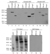
Pbx1 and Prep1 Interact with Smad2, Smad3, and Smad4 GST interaction assays were performed using the indicated bacterially expressed Smad-GST fusion proteins and 35S-labeled in vitro translated GFP, Pbx1a, Prep1, and Smad4. One tenth of the protein input and the GST tag-alone negative control are shown in lanes 13–19.
Fig. 8
Fig. 8
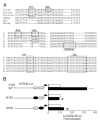
The Evolutionarily Conserved Proximal Activin Response Elements Are Also Required for Activin Regulation of the mFSHβ Gene A, Sequence comparison of activin response elements and SBEs in the ovine, bovine, porcine, human, rat, and mouse FSHβ genes. Homology is indicated by the boxed regions. B, Reporter plasmids containing either the wild-type (WT) −1000 region of the mFSHβ gene or the indicated 2-bp mutations corresponding to those made in the −167 and −134 sites of the ovine gene were transiently transfected in LβT2 cells and activin responsiveness was assayed. Data are the means ± SEM of three independent experiments, each performed in triplicate, and were normalized as described in Fig. 1. *, Values that differ significantly from vehicle-treated control; #, fold activation with activin treatment that differs significantly from fold activation of the wild-type reporter. RLU, Relative light units.
Fig. 9
Fig. 9
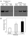
Pbx1 and Prep1 Bind to the mFSHβ Gene in Vivo Chromatin immunoprecipitation was performed using cross-linked protein/chromatin from LβT2 cells and antibodies directed against Pbx1 and Prep1 proteins. PCR primers complementary to the mFSHβ 5′-regulatory region flanking the Pbx1/Prep1 binding site were used to detect precipitation of the genomic DNA. A, Representative gel of radioactively labeled PCR products amplified from control chromatin sample and chromatin precipitated with Pbx1 and Prep1 antibodies is shown. PCR products from reactions using input chromatin at 10-fold, 50-fold, and 500-fold dilutions are shown. B, Densitometry quantification of PCR products was performed. Data are the means ± SEM from three independently prepared chromatin samples, each performed in triplicate. *, Mean densitometry values that differ significantly from no-antibody control.

References

    1. Ling N, Ying S-Y, Ueno N, Shimasaki S, Esch F, Hotta M, Guillemin R. Pituitary FSH is released by a heterodimer of the β-subunits from the two forms of inhibin. Nature. 1986;321:779–782. - PubMed
    1. Carroll RS, Corrigan AZ, Gharib SD, Vale W, Chin WW. Inhibin, activin, and follistatin: regulation of follicle-stimulating hormone messenger ribonucleic acid levels. Mol Endocrinol. 1989;3:1969–1976. - PubMed
    1. Carroll RS, Kowash PM, Lofgren JA, Schwall RH, Chin WW. In vivo regulation of FSH synthesis by inhibin and activin. Endocrinology. 1991;129:3299–3304. - PubMed
    1. Matzuk MM, Kumar TR, Bradley A. Different phenotypes for mice deficient in either activins or activin receptor type II. Nature. 1995;374:356–560. - PubMed
    1. Attisano L, Wrana JL. Signal transduction by the TGF-β superfamily. Science. 2002;296:1646–1647. - PubMed

Publication types