. 2004 Feb 17;101(7):2162-6.
doi: 10.1073/pnas.0303842101. Epub 2004 Feb 5.

LacZ-expressing olfactory ensheathing cells do not associate with myelinated axons after implantation into the compressed spinal cord

Affiliations

LacZ-expressing olfactory ensheathing cells do not associate with myelinated axons after implantation into the compressed spinal cord

J G Boyd et al. Proc Natl Acad Sci U S A. .

Abstract

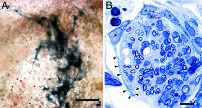
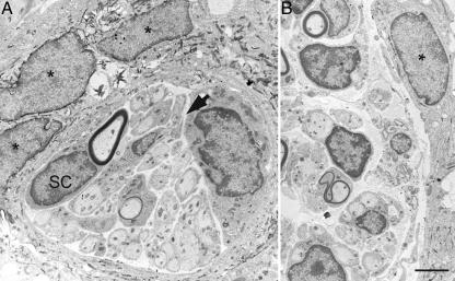
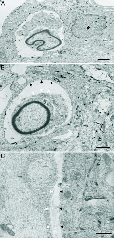
Studies have shown that implanting olfactory ensheathing cells (OECs) may be a promising therapeutic strategy to promote functional recovery after spinal cord injury. Several fundamental questions remain, however, regarding their in vivo interactions in the damaged spinal cord. We have induced a clip compression injury at the T10 level of the spinal cord in adult rats. After a delay of 1 week, OECs isolated from embryonic day 18 rats were implanted into the cystic cavity that had formed at the site of injury. Before implantation, OECs were infected with a LacZ-expressing retrovirus. At 3 weeks after implantation, LacZ-expressing OECs survived the implantation procedure and remained localized to the cystic cavity. At the electron microscopic level, the cystic cavity had clusters of LacZ-expressing OECs and numerous Schwann cells lacking LacZ expression. Although labeled OECs made no direct contact with axons, unlabeled Schwann cells were associated with either a single myelinated axon or multiple unmyelinated axons. Positively labeled OEC processes often enveloped multiple Schwann cell-axon units. These observations suggest that the role of OECs as the primary mediators of the beneficial effects on axon growth, myelination, and functional recovery after spinal cord injury may require re-evaluation.


Figures

Fig. 1.
Fig. 1.
Implantation of fetal rat OECs is associated with a robust growth of GAP-43 immunopositive processes. (A) GAP-43 immunoreactivity around the perimeter of the cystic cavity was sparse in rats that received vehicle injections. (B) Many GAP-43 immunopositive fibers were observed extending into the cystic cavity in animals that received intraspinal implantation of OECs. Low (C) and high (D) magnification photomicrographs illustrating that many of the GAP-43 immunopositive fibers (red) coexpressed neurofilament (NF; blue). (Scale bars: 400 μm, A and B; 100 μm, C; 50 μm, D.)
Fig. 2.
Fig. 2.
LacZ-expressing OECs are readily identified after intraspinal implantation. (A) A cluster of cells containing Bluo-gal reaction product was detectable within the cystic cavity 3 weeks after intraspinal implantation of LacZ-expressing OECs. The positively labeled cells did not appear to significantly migrate away from the injury site. (B) Toluidine blue-stained semithin section of OECs (*) containing blue crystalline reaction product wrapping long processes (arrowheads) around a large channel of unlabeled cells, including glia, myelinated axons, and endothelial cells. (Scale bars: 100 μm, A; 10 μm, B.)
Fig. 3.
Fig. 3.
LacZ-expressing fetal rat OECs can be identified ultrastructurally and formed distinct associations with Schwann cells in the cystic cavity. OECs containing the electron dense Bluo-gal reaction product (*) localizing to the nuclear membrane and cytoplasm were observed extending processes to envelop multiple Schwann cells (SC). These Schwann cells were associated with either large myelinated axons or several small unmyelinated axons. No Bluo-gal reaction product was detected in those glial cells associated with axons. The “tunnels” of Schwann cells are surrounded by a thin rim of collagen containing extracellular space (arrow). (Scale bar: 2.5 μm.)
Fig. 4.
Fig. 4.
LacZ-expressing OECs do not associate with axons, nor do they form a basal lamina. (A) LacZ-expressing fetal rat OECs containing the Bluo-gal reaction product did not interact directly with either myelinated or unmyelinated axons in vivo but were closely associated with Schwann cells in the cystic cavity. (B and C) LacZ-expressing OECs form tunnels in which unlabeled Schwann cells can be found within the cystic cavity. These unlabeled Schwann cells that associated with axons in vivo expressed a distinct basal lamina (white arrowheads), whereas LacZ-expressing OECs did not synthesize a basal lamina (black arrowheads). (Scale bars: 2 μm, A and B; 0.5 μm, C.)

References

    1. Sekon, L. H. S. & Fehlings, M. G. (2001) Spine 26, S2-S12. - PubMed
    1. Kwon, B. K. & Tetzlaff, W. (2001) Spine 26, S13-S22. - PubMed
    1. Bunge, M. B. (2001) Neuroscientist 7, 325-339. - PubMed
    1. Schwab, M. E. (2002) Science 295, 1029-1031. - PubMed
    1. Boyd, J. G., Skihar, V., Kawaja, M. D. & Doucette, R. (2003) Anat. Rec. 271, 49-60. - PubMed

Publication types