. 2004 Feb 17;101(7):2040-5.
doi: 10.1073/pnas.0307301101. Epub 2004 Feb 5.

Protection against electrophile and oxidant stress by induction of the phase 2 response: fate of cysteines of the Keap1 sensor modified by inducers

Affiliations

Protection against electrophile and oxidant stress by induction of the phase 2 response: fate of cysteines of the Keap1 sensor modified by inducers

Nobunao Wakabayashi et al. Proc Natl Acad Sci U S A. .

Abstract

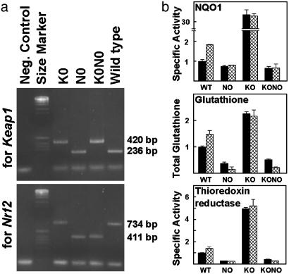
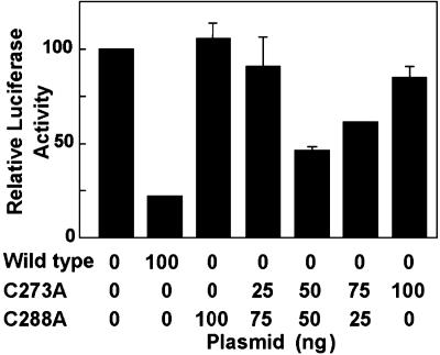
Induction of a family of phase 2 genes encoding for proteins that protect against the damage of electrophiles and reactive oxygen intermediates is potentially a major strategy for reducing the risk of cancer and chronic degenerative diseases. Many phase 2 genes are regulated by upstream antioxidant response elements (ARE) that are targets of the leucine zipper transcription factor Nrf2. Under basal conditions, Nrf2 resides mainly in the cytoplasm bound to its cysteine-rich, Kelch domain-containing partner Keap1, which is itself anchored to the actin cytoskeleton and represses Nrf2 activity. Inducers disrupt the Keap1-Nrf2 complex by modifying two (C273 and C288) of the 25 cysteine residues of Keap1. The critical role of C273 and C288 was established by (i) their high reactivity when purified recombinant Keap1 was treated with dexamethasone mesylate and the dexamethasone-modified tryptic peptides were analyzed by mass spectrometry, and (ii) transfection of keap1 and nrf2 gene-deficient mouse embryonic fibroblasts with constructs expressing cysteine to alanine mutants of Keap1, and measurement of the ability of cotransfected Nrf2 to repress an ARE-luciferase reporter. Reaction of Keap1 with inducers results in formation of intermolecular disulfide bridges, probably between C273 of one Keap1 molecule and C288 of a second. Evidence for formation of such dimers was obtained by 2D PAGE of extracts of cells treated with inducers, and by the demonstration that whereas C273A and C288A mutants of Keap1 alone could not repress Nrf2 activation of the ARE-luciferase reporter, an equal mixture of these mutant constructs restored repressor activity.


Figures

Fig. 1.
Fig. 1.
Amino acid sequence of the five domains of Keap1: (i) N-terminal region (NTR, amino acids 1–60: two cysteines, blue). (ii) BTB (Broad complex, Tramtrack, Bric-a-Brac; amino acids 61–179: three cysteines, pink), an evolutionarily conserved protein–protein interaction motif that often dimerizes with other BTB domains (33). (iii) IVR (amino acids 180–314: eight cysteines, yellow). (iv) Double glycine (DGR, amino acids 315–598: nine cysteines, gray) comprising six Kelch motifs (amino acids 315–359, 361–410, 412–457, 459–504, 506–551, and 553–598). Repeated Kelch motifs give rise to a β-propeller structure with multiple protein-binding sites (34). The DGR of Keap1 binds tightly to the Neh2 segment (the 100 N-terminal amino acids) of Nrf2 (24, 27), and is also the region involved in anchoring Keap1 to the actin cytoskeleton (19). (v) C-terminal region (CTR, amino acids 599–624: three cysteines, green). The 25 cysteine residues are highlighted in bright yellow, the 39 arginine residues are blue, and the 17 lysine residues are red. The tryptic peptides labeled with dexamethasone (T-22, T-26, T-28, T-29, and T-55) are designated. The matrix-assisted laser desorption ionization/time-of-flight mass spectral analyses of all 57 tryptic peptides, including the 19 peptides that contain cysteine residues are recorded in Table 1 (see Supporting Text).
Fig. 4.
Fig. 4.
Repression of the intensity of luciferase luminescence of ARE-luciferase in K0N0 mouse embryonic fibroblasts by wild-type Keap1 and its cysteine mutants. All cells were transfected with the ARE-luciferase (2 μg) and the Nrf2 (8 μg) constructs. (a) Repression of luminescence as a function of amount of wild-type Keap1 (black bars) and its reversal by exposure to 2 μM sulforaphane (gray bar). (b) Repression of luminescence by wild-type (WT) Keap1 (2 μg) and equivalent quantities of the following mutants: NTR (C23A and C38A); CTR (C613A, C622A, and C624A); IVR C226A–C297A (C226A, C241A, C249A, C257A, C273A, C288A, and C297A); IVR C226A–C249A (C226A, C241A, and C249A); and IVR C257A–C297A (C257A, C273A, C288A, and C297A). (c) Repressive activity of single or multiple cysteine mutants of Keap1 with 100 ng of each expression vector. The structures of the mutants are designated: + for cysteine, – for alanine. The structure of each wild-type and mutant Keap1 is shown below the bar indicating its repressor activity. Repressor activity is abrogated if C273 or C288, or both, are mutated to alanine.
Fig. 2.
Fig. 2.
Comparison of binding of wild-type and mutant (C257A–C297A) Keap1 to the Neh2 domain of Nrf2. (a) Native gel electrophoresis showing complex between Keap1 (100 pmol) and the Neh2 domain of Nrf2 (10, 20, 40, and 80 pmol, lanes 4–7 and 8–11, respectively). Lane 1, Neh2; lane 2, wild-type Keap1; lane 3, mutant Keap1. Lanes 4–7 show progressively increasing quantities of complex between wild-type Keap1 and Neh2; lanes 8–11 show lower quantities of complex formation between mutant Keap1 and Neh2. Arrowhead, arrow, and asterisk show Keap1, Neh2, and Neh2-Keap1 or mutant Keap1 complex bands, respectively. (b) Densitometric quantification of the intensities of bands of complexes between Neh2 and wild-type Keap1 (black bars) and mutant Keap1 (hatched bars).
Fig. 3.
Fig. 3.
Genotyping and levels of expression of phase 2 gene products in established lines of embryonic fibroblasts obtained from mice in which the keap1 (K0), nrf 2 (N0), or both genes (K0N0) were disrupted. (a) Electrophoresis of the PCR products derived from keap1 wild-type (236 bp) and mutant (420 bp) alleles, and nrf2 wild-type (734 bp) and mutant (411 bp) alleles. (b) Comparison (normalized to wild-type controls) of the specific activities of NQO1 and thioredoxin reductase, and concentration of glutathione in cell-free extracts of the three mutant and wild-type mouse embryonic fibroblasts. Black bars, cells untreated with inducers; hatched bars, cells exposed for 24 h to 1.5 μM sulforaphane.
Fig. 5.
Fig. 5.
Repression of the intensity of luciferase luminescence of ARE-luciferase in K0N0 mouse embryo fibroblasts by wild-type Keap1 and its C273A and C288A mutants. All cells were transfected with the ARE-luciferase (2 μg) and the Nrf2 (8 μg) constructs. The luminescence observed in the absence of Keap1 is expressed as 100 units. Addition of 100 ng of wild-type Keap1 plasmid reduced the luminescence to ≈20%. The plasmids (100 ng) coding for C273A or C288A showed little, if any, repression of the fluorescence, whereas mixtures of these plasmids restored the repressor activity. A mixture of 50 ng of each plasmid repressed ≈40% of the luminescence of the system.
Fig. 6.
Fig. 6.
Exposure to inducers causes formation of a disulfide-linked dimer of Keap1 in HEK 293 cells transfected with a construct encoding for Keap1 and GFP (normalization control). (a) Coomassie brilliant blue (CBB) staining of 2D SDS/PAGE of cell-free extracts. (b) Immunoblots of SDS/PAGE of control (lanes 1 and 2) and inducer-treated (lanes 3 and 4) cells showing (Top) reduced binding of the anti-Keap1 antibody for Keap1 in inducer-treated cells compared with control cells, (Middle) equal expression of GFP, and (Bottom) equal cell numbers as judged by the expression of Lamin B. (c) Immunoblots for Keap1 of 2D SDS/PAGE of extracts of control cells and cells exposed to inducers of three different chemical types. SF, sulforaphane; D3T, 1,2-dithiole-3-thione; 2-HBA, bis(2-hydroxybenzylidene)acetone.
Fig. 7.
Fig. 7.
Mechanism of regulation of the phase 2 response. Nrf2 (black) is retained in the cytoplasm by interaction with two molecules of Keap1, which are dimerized through their BTB domains (pink) and anchored to the actin cytoskeleton via the Kelch or DGR region (gray propeller). Inducers of the phase 2 response interact with cysteine thiol groups in the intervening region (IVR, yellow) of Keap1, causing the formation of disulfide bonds (most likely between C273 of one monomer and C288 of the other). This results in conformational change that renders Keap1 unable to bind to Nrf2, which then translocates to the nucleus. The Nrf2 in heterodimeric combination with other transcription factors such as small Maf binds to the ARE regulatory region of phase 2 genes and enhances their transcription.

References

    1. Hayes, J. D. & McLellan, L. I. (1999) Free Radical Res. 31, 273–300. - PubMed
    1. Talalay, P., Dinkova-Kostova, A. T. & Holtzclaw, W. D. (2003) Adv. Enzyme Regul. 43, 121–134. - PubMed
    1. Talalay, P. (1999) Proc. Am. Philos. Soc. 143, 52–72.
    1. Talalay, P. (2000) Biofactors 12, 5–11. - PubMed
    1. Ramos-Gomez, M., Kwak, M. K., Dolan, P. M., Itoh, K., Yamamoto, M., Talalay, P. & Kensler, T. W. (2001) Proc. Natl. Acad. Sci. USA 98, 3410–3415. - PMC - PubMed

Publication types

MeSH terms

LinkOut - more resources