. 2004 Feb 17;101(7):2076-81.
doi: 10.1073/pnas.0308334100. Epub 2004 Feb 5.

The role of Shc and insulin-like growth factor 1 receptor in mediating the translocation of estrogen receptor alpha to the plasma membrane

Affiliations

The role of Shc and insulin-like growth factor 1 receptor in mediating the translocation of estrogen receptor alpha to the plasma membrane

Robert X Song et al. Proc Natl Acad Sci U S A. .

Abstract

Our previous studies demonstrated that 17beta-estradiol (E2) rapidly induces the interaction of estrogen receptor alpha (ERalpha) with the adapter protein Shc, the translocation of ERalpha to the cell membrane, and the formation of dynamic membrane structures in MCF-7 breast cancer cells. The present study examined how E2 causes ERalpha to translocate to the region of the plasma membrane and focused on mechanisms whereby Shc and the insulin-like growth factor-1 receptor (IGF-1R) mediate this process. Shc physically interacts with IGF-1R in the plasma membrane, and E2 activates IGF-1R. We reasoned that ERalpha, when bound to Shc, would be directed to the region of the plasma membrane by the same processes, causing membrane translocation of Shc. We confirmed that E2 rapidly induced IGF-1R phosphorylation and demonstrated that E2 induced formation of a ternary protein complex among Shc, ERalpha, and IGF-1R. Knock down of Shc with a specific small inhibitory RNA decreased the association of ERalpha with IGF-1R by 87%, suggesting that Shc is a crucial molecule in the formation of this ternary complex. Confocal microscopy studies provided further confirmation of the functional roles of Shc and the IGF-1R in the translocation of ERalpha to the region of the membrane. Down-regulation of Shc, ERalpha, or IGF-1R with specific small inhibitory RNAs all blocked E2-induced mitogen-activated protein kinase phosphorylation. Together, our results demonstrate that Shc and IGF-1R serve as key elements in the translocation of ERalpha to the cell membrane and in the facilitation of ERalpha-mediated rapid E2 action.


Figures

Fig. 1.
Fig. 1.
(A) E2-induced protein complex formation among ERα, Shc, and IGF-1R. MCF-7 cells were treated with vehicle, 1 ng/ml IGF-1, or E2 at 0.1 nM for the times indicated. Lysates were immunoprecipitated with IGF-1R antibody. The nonspecific monoclonal antibody (IgG) served as a negative control. (B) The effect of ICI on E2-induced protein-protein interaction was assayed by immunoprecipitation of IGF-1R and detection of ERα on Western blot.
Fig. 2.
Fig. 2.
Protein silencing and the role of Shc in protein complex formation in MCF-7 cells. (A) Cells were transfected with nonspecific or specific siRNA for 2 days and then treated with vehicle or 0.1 nM E2 for 15 min for additional MAPK phosphorylation assay. Polyvinylidene difluoride membranes were probed with the specific antibodies to detect the protein expression of IGF-1R, ERα, and Shc. (B) Confocal microscopy study of siRNA expression in MCF-7 cells. Cells, transfected with or without siRNA against Shc, were grown on cover slips and then subjected to immunofluorescence staining for Shc (blue) and ERα (green), as described in Materials and Methods. (C and D) The role of Shc in mediating the interaction of ERα and IGF-1R. Cells transfected with siRNA as indicated were treated with vehicle or E2 at 0.1 nM. The IGF-1R and ERα interaction was assayed by immunoprecipitation of IGF-1R and detection of ERα on Western blot (C) or by immunoprecipitation of ERα and detection of IGF-1R on Western blot (D). The data are from three experiments combined, and only one experiment is shown. *, P < 0.05 comparison of E2-treated cells with vehicle control.
Fig. 3.
Fig. 3.
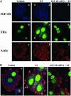
Shc involvement in E2-induced ERα membrane association in MCF-7 cells. Cells were transfected with or without siRNA directed against Shc. Two days later, cells were treated with vehicle or E2 (0.1 nM) for 15 min and then subjected to immunofluorescence staining with anti-Shc anti-ERα antibodies. Actin was stained with phalloidin, indicating filamentous cell membrane. (A) The nonmerged images show Shc in blue, ERα in green, and cell membrane in red. (Left) The vehicle-treated cells stained with Shc, ERα, and actin. (Center and Right) Estrogen-treated cells with or without siRNA of Shc expression. (B) Colocalization of ERα, Shc, and actin on cell membrane is shown in three color-merged images. To highlight the specific changes, the arrows illustrate one example of the effects in single cells. Close inspection reveals multiple changes not marked by arrows.
Fig. 4.
Fig. 4.
IGF-1R involvement in E2-induced ERα membrane association in MCF-7 cells. Cells cultured on coverslips were transfected with or without siRNA against IGF-1R for 2 days and then treated with vehicle or E2 at 0.1 nM for 15 min. After fixation, cells were immunofluorescently stained with anti-IGF-1R (blue) anti-ERα (green) antibodies. Actin was stained with phalloidin (red), indicating filamentous cell membrane. Both nonmerged (A) and merged (B) images are shown.
Fig. 5.
Fig. 5.
Evidence of the involvement of ERα, Shc, and IGF-1R in E2-induced MAPK phosphorylation. (A) Down-regulation of ERα, Shc, and IGF-1R blocked E2-induced MAPK phosphorylation. Cells were transfected with specific and nonspecific siRNAs. At day 2, cells were treated with vehicle or E2 at 0.1 nM for 15 min, and then whole-cell extracts were prepared and analyzed by Western blotting as described in Materials and Methods. Polyvinylidene difluoride membranes were probed with antidual phosphotyrosine MAPK antibody. *, P < 0.05 comparison of E2-treated cells with the vehicle-treated control in each siRNA transfected group. (B) Selective pathway inhibitors on E2-induced MAPK phosphorylation. Cells were challenged with vehicle or 0.1 nM E2 for 15 min in the presence of the inhibitors, as indicated.

Similar articles

Cited by

References

    1. Yager, J. D. (2000) J. Natl. Cancer Inst. Monogr. 27, 67-73. - PubMed
    1. Pietras, R. J., Nemere, I. & Szego, C. M. (2001) Endocrine 14, 417-427. - PubMed
    1. Ho, K. J. & Liao, J. K. (2002) Arterioscler. Thromb. Vasc. Biol. 22, 1952-1961. - PubMed
    1. Collins, P. & Webb, C. (1999) Nat. Med. 5, 1130-1131. - PubMed
    1. Watson, C. S., Campbell, C. H. & Gametchu, B. (1999) Exp. Physiol. 84, 1013-1022. - PMC - PubMed

Publication types

MeSH terms

Substances

LinkOut - more resources