

Expansion and contraction of the hepatitis B virus transcriptional template in infected chimpanzees
- PMID: 14764900
- PMCID: PMC357063
- DOI: 10.1073/pnas.0308478100
Expansion and contraction of the hepatitis B virus transcriptional template in infected chimpanzees
Abstract
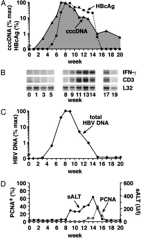
We have previously shown that hepatitis B virus (HBV) replication is controlled by noncytolytic mechanisms that depend primarily on the effector functions of the CD8(+) T cell response, especially the production of IFN-gamma in the liver. The mechanisms that control the nuclear pool of viral covalently closed circular DNA (cccDNA) transcriptional template of HBV, which must be eliminated to eradicate infection, have been difficult to resolve. To examine those mechanisms, we quantitated intrahepatic HBV cccDNA levels in acutely infected chimpanzees whose virological, immunological, and pathological features were previously described. Our results demonstrate that the elimination kinetics of the cccDNA are more rapid than the elimination of HBV antigen-positive hepatocytes during the early phase of viral clearance, and they coincide with the influx of small numbers of IFN-gamma producing CD8(+) T cells into the liver. In contrast, terminal clearance of the cccDNA is associated with the peak of liver disease and hepatocellular turnover and with a surge of IFN-gamma producing CD8(+) T cells in the liver. Collectively, these results suggest that cccDNA clearance is a two-step process mediated by the cellular immune response. The first step reduces the pool of cccDNA molecules noncytolytically, probably by eliminating their relaxed circular DNA precursors and perhaps by destabilizing them. The second step enhances this process by destroying infected hepatocytes and triggering their turnover. Surprisingly, despite this multipronged response, traces of cccDNA persist indefinitely in the liver, likely providing a continuous antigenic stimulus that confers lifelong immunity.
Figures


Similar articles
-
Interferon-γ and Tumor Necrosis Factor-α Produced by T Cells Reduce the HBV Persistence Form, cccDNA, Without Cytolysis.Gastroenterology. 2016 Jan;150(1):194-205. doi: 10.1053/j.gastro.2015.09.026. Epub 2015 Sep 28. Gastroenterology. 2016. PMID: 26416327
-
In vivo proliferation of hepadnavirus-infected hepatocytes induces loss of covalently closed circular DNA in mice.Hepatology. 2010 Jul;52(1):16-24. doi: 10.1002/hep.23611. Hepatology. 2010. PMID: 20578126
-
Viral clearance without destruction of infected cells during acute HBV infection.Science. 1999 Apr 30;284(5415):825-9. doi: 10.1126/science.284.5415.825. Science. 1999. PMID: 10221919
-
Attacking hepatitis B virus cccDNA--The holy grail to hepatitis B cure.J Hepatol. 2016 Apr;64(1 Suppl):S41-S48. doi: 10.1016/j.jhep.2016.02.009. J Hepatol. 2016. PMID: 27084036 Review.
-
Revisiting Hepatitis B Virus: Challenges of Curative Therapies.J Virol. 2019 Sep 30;93(20):e01032-19. doi: 10.1128/JVI.01032-19. Print 2019 Oct 15. J Virol. 2019. PMID: 31375584 Free PMC article. Review.
Cited by
-
GS-9620, an oral agonist of Toll-like receptor-7, induces prolonged suppression of hepatitis B virus in chronically infected chimpanzees.Gastroenterology. 2013 Jun;144(7):1508-17, 1517.e1-10. doi: 10.1053/j.gastro.2013.02.003. Epub 2013 Feb 13. Gastroenterology. 2013. PMID: 23415804 Free PMC article.
-
Metabolism and function of hepatitis B virus cccDNA: Implications for the development of cccDNA-targeting antiviral therapeutics.Antiviral Res. 2015 Oct;122:91-100. doi: 10.1016/j.antiviral.2015.08.005. Epub 2015 Aug 10. Antiviral Res. 2015. PMID: 26272257 Free PMC article. Review.
-
Human liver chimeric mice provide a model for hepatitis B and C virus infection and treatment.J Clin Invest. 2010 Mar;120(3):924-30. doi: 10.1172/JCI40094. Epub 2010 Feb 22. J Clin Invest. 2010. PMID: 20179355 Free PMC article.
-
Genomic analysis of the host response to hepatitis B virus infection.Proc Natl Acad Sci U S A. 2004 Apr 27;101(17):6669-74. doi: 10.1073/pnas.0401771101. Epub 2004 Apr 20. Proc Natl Acad Sci U S A. 2004. PMID: 15100412 Free PMC article.
-
An age-structured model of hepatitis B viral infection highlights the potential of different therapeutic strategies.Sci Rep. 2022 Jan 24;12(1):1252. doi: 10.1038/s41598-021-04022-z. Sci Rep. 2022. PMID: 35075156 Free PMC article.
References
Publication types
MeSH terms
Substances
Grants and funding
LinkOut - more resources
Full Text Sources
Medical
Research Materials
