. 2004 Feb 25;23(4):919-29.
doi: 10.1038/sj.emboj.7600103. Epub 2004 Feb 12.

Fission yeast Clp1p phosphatase affects G2/M transition and mitotic exit through Cdc25p inactivation

Affiliations

Fission yeast Clp1p phosphatase affects G2/M transition and mitotic exit through Cdc25p inactivation

Benjamin A Wolfe et al. EMBO J. .

Abstract

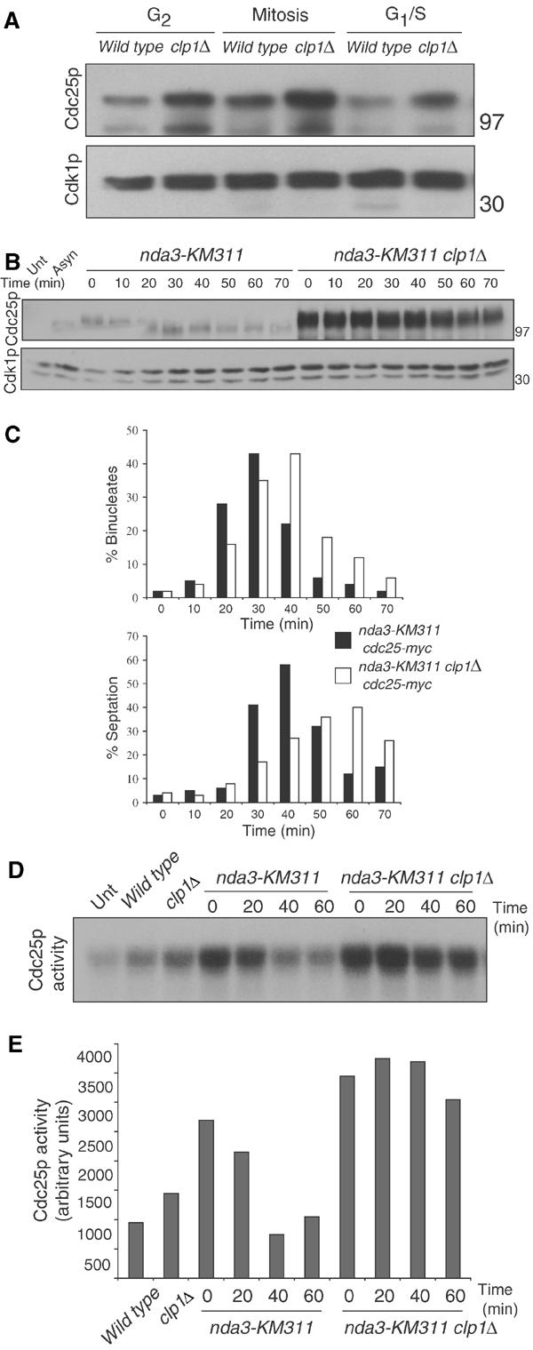
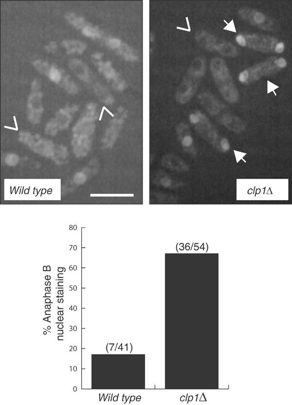
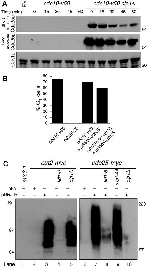
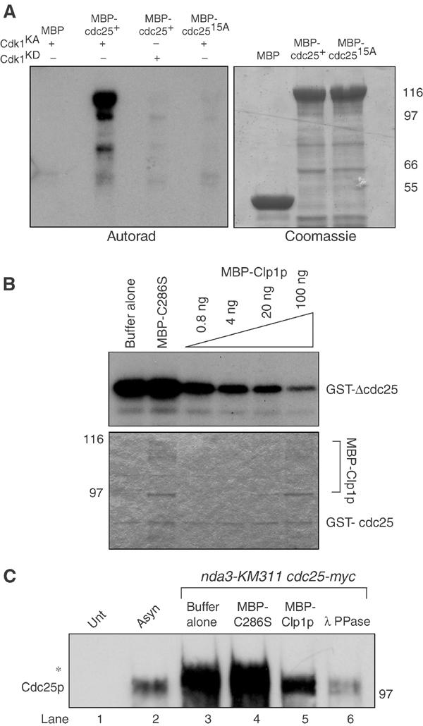
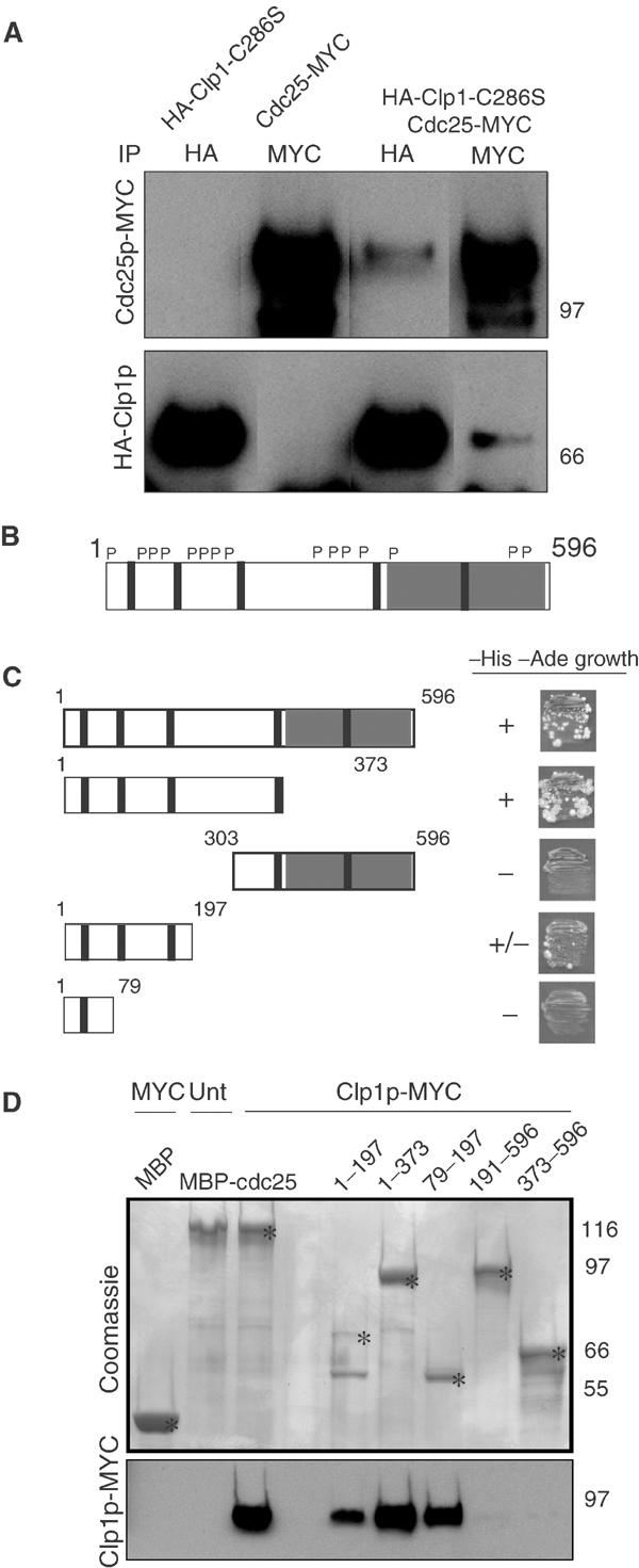
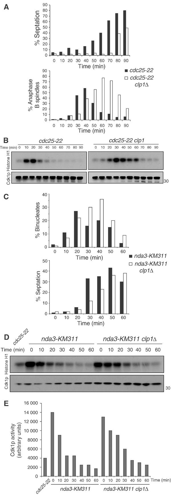
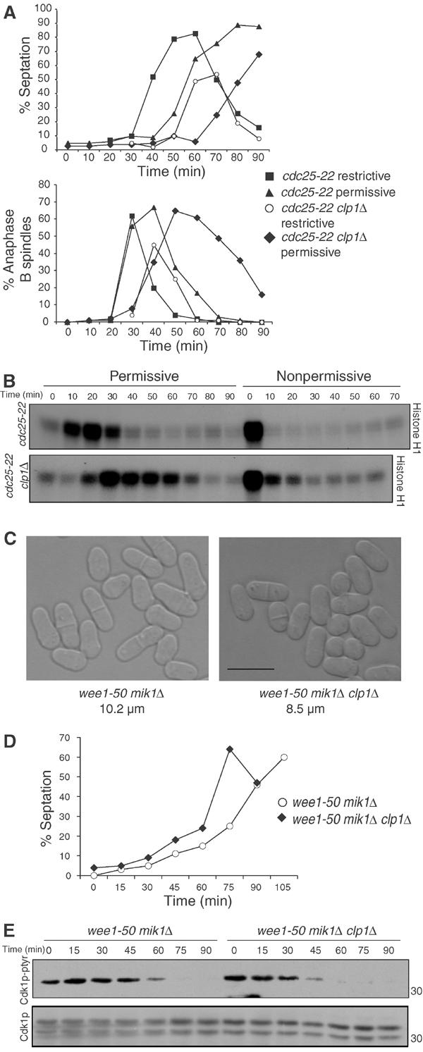
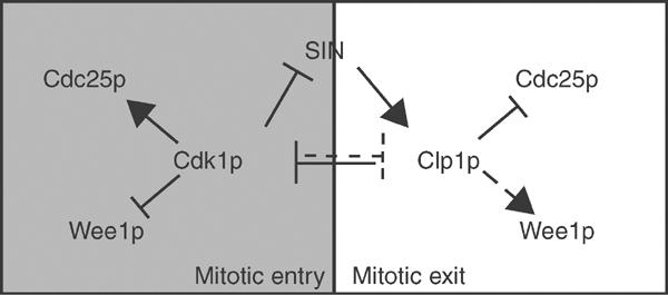
The Cdc14 family of phosphatases specifically reverses proline-directed phosphorylation events. In Saccharomyces cerevisiae, Cdc14p promotes Cdk1p inactivation at mitotic exit by reversing Cdk1p-dependent phosphorylations. Cdk1p is a proline-directed kinase whose activity is required in all eukaryotes for the transit into mitosis. At mitotic commitment, Cdk1p participates in its own regulation by activating the mitotic inducing phosphatase, Cdc25p, and inhibiting the opposing kinase, Wee1p. We have investigated the ability of Schizosaccharomyces pombe Clp1p, a Cdc14p homolog, to disrupt this auto-amplification loop. We show here that Clp1p is required to dephosphorylate, destabilize, and inactivate Cdc25p at the end of mitosis. Clp1p promotes recognition of Cdc25p by the anaphase-promoting complex/cyclosome, an E3 ubiquitin ligase. Failure to inactivate and destabilize Cdc25p in late mitosis delays progression through anaphase, interferes with septation initiation network signaling, and additionally advances the commitment to mitotic entry in the next cycle. This may be a widely conserved mechanism whereby Cdc14 proteins contribute to Cdk1p inactivation.


Figures

Figure 1
Figure 1
clp1+ regulation of Cdc25p. (A) cdc25-myc (KGY 3377) or cdc25-myc clp1Δ (KGY 3499) were grown to mid-log phase and synchronized by centrifugal elutriation. Samples were taken after synchronization and processed for protein (A) and cell cycle stage by DAPI staining and cell measurement analysis as follows: G2 cells were uninucleate and 8–12 μm in length, mitotic cells were uninucleate or binucleate cells with condensed chromatin, and G1/S cells contained two interphase nuclei and a septum (data not shown). Cdc25p-myc and Cdk1p (serves as loading control in each of our blots) were detected by immunoblotting with 9E10 and anti-PSTAIR antibodies, respectively. Numbers adjacent to the blots indicate the position of molecular weight standards on this and all subsequent blots. (B) nda3-KM311 cdc25-myc (KGY 3916) and nda3-KM311 cdc25-myc clp1Δ (KGY 4213) were grown to mid-log phase and synchronized by cold shift to 18°C for 7 h. Cells were then released to the permissive temperature (32°C), and samples were collected at the indicated time points. Extracts were prepared from these and processed for immunoblot analysis. (C) Completion of mitotic exit was monitored by determining binucleate formation (top panel) and septation index (lower panel). For the Cdc25p immunoblot, samples were immunoprecipitated with 9E10 antibody and immunoblotted with anti-Cdc25p antibodies. (D) Cells were synchronized as in (B) and samples were collected at the indicated time points. Extracts were prepared and immunoprecipitated with 9E10 antibody. Samples were processed for Cdc25p activity and quantified and normalized to basal levels of activity (Unt) in (E) indirectly via activation of Cdk1p's histone H1 activity as described in Materials and methods. In this experiment, Cdc25p activity was a saturable reaction (data not shown).
Figure 2
Figure 2
Cdc25p remains nuclear in clp1Δ anaphase B cells. (A) Compression of image stacks of cdc25-GFP (KGY 4337) and cdc25-GFP clp1Δ (KGY 4341) asynchronous cells grown at 25°C in YE medium. Tail-less arrows (∨) indicate binucleate cells without Cdc25p-GFP nuclear fluorescence and arrows (↓) indicate binucleate cells with nuclear Cdc25p-GFP. Scale bar indicates 10 μm. (B) Percentage of anaphase B cells with nuclear Cdc25p-GFP from cdc25-GFP sid4-GFP (KGY 877) and cdc25-GFP sid4-GFP clp1Δ (KGY 148) asynchronous cells grown at 25°C in YE medium. Anaphase B cells were those with two well-separated spindle pole bodies (Sid4p-GFP) and no septum.
Figure 3
Figure 3
Cdc25p destabilization and ubiquitination requires Clp1p and APC/C. (A) cdc10-V50 (KGY 1744) and cdc10-V50 clp1Δ (KGY 2752) cultures expressing pREP41myc-cdc25C480S were grown in the absence of thiamine for 20 h at the permissive temperature (25°C), and then shifted to the nonpermissive temperature (36°C) for 3.5 h, at which point excess thiamine (4 μM) and cyclohexamide (100 μg/ml) were added to the cultures. Samples were taken at the indicated time points. Extracts were prepared and immunoblotted with 9E10 and anti-PSTAIR to detect Cdc25p and Cdk1p, respectively. (B) Percentage of G1 cells from (A) as determined by Sytox Green (Molecular Probes) staining and flow cytometry. (C) In vivo ubiquitination assays. mts3-1 (KGY 574) (lane 1), mts3-1 cut2-myc (KGY 1923) (lane 3), mts3-1 cut2-myc lid1-6 (KGY 1948) (lane 4), mts3-1 cut2-myc clp1Δ (KGY 878) (lane 5), mts3-1 cdc25-myc (KGY 4366) (lane 7), mts3-1 lid1-6 cdc25-myc (KGY 100) (lane 8), mts3-1 skp1-A4 cdc25-myc (KGY 102) (lane 9), and mts3-1 clp1Δ cdc25-myc (KGY 110) (lane 10) strains transformed with pREP1-His6-ubiquitin, and mts3-1 cut2-myc (KGY 1923) (lane 2) and mts3-1 cdc25-myc (KGY 4366) (lane 6) transformed with empty vector were grown at 25°C for 22 h in the absence of thiamine to induce His6-ubiquitin expression and then shifted to 36°C for an additional 4 h. Samples were collected and extracts were prepared. Ubiquitin conjugates were purified on Ni-NTA beads, separated on SDS–PAGE, and immunoblotted with 9E10 antibodies to detect Cut2p and Cdc25p.
Figure 4
Figure 4
Clp1p reverses Cdk1p-dependent phosphorylation of Cdc25p. (A) Approximately 1–2 μg of MBP, MBP-Cdc25p, or MBP-Cdc25p-15A was phosphorylated in vitro with baculoviral produced and purified recombinant active (KA) or kinase dead (KD) Cdk1p complex. Reactions were separated by SDS–PAGE and analyzed by Coomassie blue staining and autoradiography. (B) Approximately 100 ng of GST-ΔCdc25p containing amino acids 1–197 was phosphorylated by recombinant Cdk1p complex in vitro as in (A), and subsequently incubated with the indicated amounts of MBP-C286S or MBP-Clp1p. Reactions were separated by SDS–PAGE, and analyzed by Coomassie blue staining (lower panel) and autoradiography (upper panel). (C) Cell pellets from nda3KM311 cdc25-myc (KGY 3916) arrested at the restrictive temperature (18°C), and wild type (KGY 246) (Unt) and cdc25-myc (KGY 3377) (Asyn) grown to mid-log phase were collected. Extracts were prepared and immunoprecipitated with 9E10 antibodies and subsequently incubated with phosphatase buffer alone (lanes 1, 2, and 3), 100 ng MBP-C286S (lane 4), 100 ng MBP-Clp1p (lane 5), or 0.5 μg Lambda phosphatase (lane 6). Reactions were separated by SDS–PAGE and immunoblotted with anti-Cdc25p antibodies. The asterisk (*) indicates the position of the hyperphosphorylated Cdc25p.
Figure 5
Figure 5
Clp1p interacts with the N-terminus of Cdc25p. (A) wild type (KGY 246) or cdc25-myc (KGY 3377) cells were transformed with empty vector or pREP41- HA-clp1C286S, and expression was induced by growth in media lacking thiamine for 18 h at 32°C. Samples were collected and extracts were prepared. Native lysates were split, immunoprecipitated with 9E10 and 12CA5 antibodies, and separated by SDS–PAGE. Membranes were probed with 9E10 and 12CA5 antibodies to detect MYC- and HA-tagged proteins, respectively. (B) Schematic representation of Cdc25p depicting carboxy terminal catalytic domain (gray box), putative Cdk1p phosphorylation sites (P), and five putative destruction boxes (∣). (C) Strain PJ206 (KGY 1296) was cotransformed with bait plasmid pGBT9:clp1 and the indicated cdc25 fragments in the prey plasmid, pGAD424, and then screened for ability (±) to support growth on –His –Ade (double selection) plates. (D) Extracts were prepared from either wild type (KGY 246) (Unt) or clp1-MYC (KGY 2882) (Clp1p-MYC) strains grown in YE media at 32°C, and incubated in the presence of the indicated MBP-Cdc25p fragments bound to amylose beads. Beads were washed extensively and samples were separated by SDS–PAGE. Proteins were either detected by Coomassie blue staining or transferred to PVDF and subsequently probed with 9E10 antibodies to detect Clp1p. Asterisks (*) indicate the position of the predicted size of the fusion protein on Coomassie-stained gel.
Figure 6
Figure 6
clp1Δ cells delay Cdk1p inactivation at the end of mitosis. cdc25-22 (KGY 851) and cdc25-22 clp1Δ (KGY 3380) cells were arrested in G2 by incubation at 36°C for 4 h. Cultures were then released to 25°C, and samples were taken at the indicated time points and fixed with ethanol or frozen. (A) Septation index was calculated using a light microscope (top panel). Ethanol-fixed samples were subjected to indirect immunofluorescence with anti-TAT1 antibodies (bottom panel). The presence of elongated spindles and separated DNA masses was assessed in at least 200 cells. (B) Cell pellets were lysed under native conditions and immunoprecipitated for Cdk1p with 4711 antibody. Samples were processed for histone H1 kinase activity and immunoblotted for Cdk1p levels with anti-PSTAIR. (C) nda3-KM311 (KGY 3612) and nda3-KM311 clp1Δ (KGY 3783) were arrested in prometaphase by incubation at 18°C for 6.5 h. Cells were released at 32°C, and ethanol-fixed samples and cell pellets were collected at the indicated time points. Septation index was determined as in (A) (C, lower panel) and fixed cells were stained with DAPI to visualize nuclei. The presence of separated DNA masses and no septum was assessed in at least 200 cells (C, top panel). (D) Cell pellets were analyzed as in (B) and Cdk1p activity was quantified (E).
Figure 7
Figure 7
Exit delay and G2 advancement depend in large part on Cdc25p. (A) cdc25-22 (KGY 851) and cdc25-22 clp1Δ (KGY 3380) were arrested in G2 by incubation at 36°C for 4 h and released to 25°C to undergo a synchronous mitosis. The timing of mitotic entry was judged by histone H1 kinase assays as well as microtubule staining for metaphase spindles in several previous experiments and confirmed in this one. After cells entered mitosis (20 min for wild type and 30 min for clp1Δ), the cultures were spilt in half, and incubated either at 25°C (permissive) or 36°C (nonpermissive). Ethanol-fixed samples and cell pellets were taken at the indicated time points. (A) Septation was determined by light microscopy (top panel). Ethanol-fixed samples were subjected to indirect immunofluorescence with anti-TAT1 antibodies (bottom panel). The presence of elongated spindles and separated DNA masses was assessed in at least 200 cells. (B) Cell pellets were lysed under native conditions and immunoprecipitated for Cdk1p with anti-4711. Immunecomplexes were processed for histone H1 kinase activity and immunoblotted for Cdk1p with anti-PSTAIR antibodies. (C) Differential interference contrast images of wee1-50 mik1Δ (KGY 612) and wee1-50 mik1Δ clp1Δ (KGY 98) grown at 25°C in YE medium. Cell lengths at septation were scored from at least 20 cells using Openlab software (Improvision). The scale bar indicates 10 μm. (D, E) wee1-50 mik1Δ (KGY 612) and wee1-50 mik1Δ clp1Δ (KGY 98) cells grown at 25°C were synchronized by centrifugal elutriation. Cells were released to the restrictive temperature (36°C), and samples taken at the indicated time points were processed for % septation (D) and for protein. (E) Lysates were prepared under native conditions and separated on SDS–PAGE. Membranes were immunoblotted for Tyr-15 phosphorylated Cdk1p and total Cdk1p with phospho-Tyr-15 Cdk1p and PSTAIR antibodies, respectively.
Figure 8
Figure 8
Positive and negative regulation of Cdk1p during mitosis. During mitosis, Cdk1p stimulates its own activity by activating its positive regulator (Cdc25p) and inactivating its negative regulators (Wee1p, SIN, Clp1p). As Cdk1p activity is sufficiently downregulated at mitotic exit, SIN and Clp1p function together to combat Cdk1p activation by reversing the auto-amplification loop and promoting cytokinesis.

References

    1. Aligue R, Wu L, Russell P (1997) Regulation of Schizosaccharomyces pombe Wee1 tyrosine kinase. J Biol Chem 272: 13320–13325 - PubMed
    1. Bahler J, Wu JQ, Longtine MS, Shah NG, McKenzie A 3rd, Steever AB, Wach A, Philippsen P, Pringle JR (1998) Heterologous modules for efficient and versatile PCR-based gene targeting in Schizosaccharomyces pombe. Yeast 14: 943–951 - PubMed
    1. Bardin AJ, Amon A (2001) Men and sin: what's the difference? Nat Rev Mol Cell Biol 2: 815–826 - PubMed
    1. Bembenek J, Yu H (2001) Regulation of the anaphase-promoting complex by the dual specificity phosphatase human Cdc14a. J Biol Chem 276: 48237–48242 - PubMed
    1. Benito J, Martin-Castellanos C, Moreno S (1998) Regulation of the G1 phase of the cell cycle by periodic stabilization and degradation of the p25rum1 CDK inhibitor. EMBO J 17: 482–497 - PMC - PubMed

Publication types

MeSH terms

LinkOut - more resources