. 2004 Feb 25;23(4):701-11.
doi: 10.1038/sj.emboj.7600100. Epub 2004 Feb 5.

The Yersinia adhesin YadA collagen-binding domain structure is a novel left-handed parallel beta-roll

Affiliations

The Yersinia adhesin YadA collagen-binding domain structure is a novel left-handed parallel beta-roll

Heli Nummelin et al. EMBO J. .

Abstract

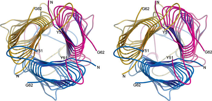
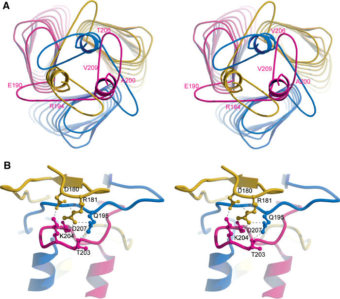
The crystal structure of the recombinant collagen-binding domain of Yersinia adhesin YadA from Yersinia enterocolitica serotype O:3 was solved at 1.55 A resolution. The trimeric structure is composed of head and neck regions, and the collagen binding head region is a novel nine-coiled left-handed parallel beta-roll. Before the beta-roll, the polypeptide loops from one monomer to the rest, and after the beta-roll the neck region does the same, making the transition from the globular head region to the narrower stalk domain. This creates an intrinsically stable 'lock nut' structure. The trimeric form of YadA is required for collagen binding, and mutagenesis of its surface residues allowed identification of a putative collagen-binding surface. Furthermore, a new structure-sequence motif for YadA beta-roll was used to identify putative YadA-head-like domains in a variety of human and plant pathogens. Such domains may therefore be a common bacterial strategy for avoiding host response.


Figures

Figure 1
Figure 1
Electron density of (2Fo-Fc) map contoured at 1.5σ for turns T1 (A) and T2 (B) showing S113 and N146 in i+2 positions in left-handed α-helical conformation. Hydrogen bonding from G111 and G144 to i+3 is illustrated in violet with the distance between atoms in Å. The figure was prepared using BOBSCRIPT (Esnouf, 1999) and Raster3D. (C) Stereo picture of trimeric YadA head domain. The N-termini are up and the C-termini are down. The strands are drawn as arrows, helices as ribbons and the different monomers are in different colours.
Figure 2
Figure 2
(A) One level of the β-roll in the trimer viewed along the z-axis showing the packing of the oligomeric core by large hydrophobic residues and the packing of the monomeric interior by small hydrophobic residues. The conserved (ii+3) hydrogen bonds are shown with dashed lines. The colouring of the monomers is the same as in Figure 1C. (B) Alignment of the full β-roll repeats. The NSVAIGXXS repeats are shown in bold and the totally conserved Gly is boxed. The turns T1 and T2 are marked on top of the alignment and the residues making β-strands are in grey boxes. The consensus sequence is marked below the alignment.
Figure 3
Figure 3
The YadA head domain viewed down the z-axis from the N-terminus. The twisting of the monomers and the extension of the coils in different directions at the top and the bottom of the LPBRs is seen. On the top of the trimer, strands 32–40 are trapped under loops 41–50. The colours indicate the most plausible connectivity of a separate N-terminal loop with the rest of the monomer. The colouring of the monomers is the same as in Figure 1C.
Figure 4
Figure 4
(A) Organisation of the neck region in the C-terminus of the head domain, viewed from the C-terminus along the z-axis. The safety-pin structures as well as the beginnings of the stalk domain helices are shown. Some residues in the neck region are numbered for the magenta monomer for clarity. (B) The neck region (viewed perpendicular to (A) showing one multi-centre ionic network in the safety-pin region. It ties the three ‘safety pins' to the central β-roll assembly. The colouring of the monomers is the same as in Figure 1C.
Figure 5
Figure 5
Most likely orientation of the collagen triple helix on the surface of the trimeric YadA head domain. The mutant residues are coloured according to their effect on type I collagen so that the mutants that abolished the binding totally are coloured red, to <20% in orange and the ones that attenuated the binding in violet. The residues D180 and E182, which also had an effect on the trimeric structure, are in blue. The figure was prepared using PyMOL (DeLano, 2002).

References

    1. Aghajari N, Van Petegem F, Villeret V, Chessa J-P, Gerday C, Haser R, Van Beeumen J (2003) Crystal structures of a psychrophilic metalloprotease reveal new insights into catalysis by cold-adapted psroteases. PROTEINS: Struct Funct Genet 50: 636–647 - PubMed
    1. Balligand G, Laroche Y, Cornelis G (1985) Genetic analysis of virulence plasmid from a serogroup 9 Yersinia enterocolitica strain: role of outer membrane protein P1 in resistance to human serum and autoagglutination. Infect Immun 48: 782–786 - PMC - PubMed
    1. Baumann U (1994) Crystal structure of the 50 kDa metallo protease from Serratia marcescens. J Mol Biol 242: 244–251 - PubMed
    1. Baumann U, Wu S, Flaherty KM, McKay DB (1993) Three-dimensional structure of the alkaline protease of Pseudomonas aeruginosa: a two-domain protein with a calcium-binding parallel beta roll motif. EMBO J 12: 3357–3364 - PMC - PubMed
    1. Berman HM, Westbrook J, Feng Z, Gilliland G, Bhat TN, Weissig H, Shindyalov IN, Bourne PE (2000) The protein data bank. Nucleic Acids Res 28: 235–242 - PMC - PubMed

Publication types

MeSH terms