. 2004 Feb 25;23(4):712-8.
doi: 10.1038/sj.emboj.7600093. Epub 2004 Feb 12.

Crystal structure of a myristoylated CAP-23/NAP-22 N-terminal domain complexed with Ca2+/calmodulin

Affiliations

Crystal structure of a myristoylated CAP-23/NAP-22 N-terminal domain complexed with Ca2+/calmodulin

Mamoru Matsubara et al. EMBO J. .

Abstract

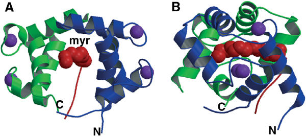
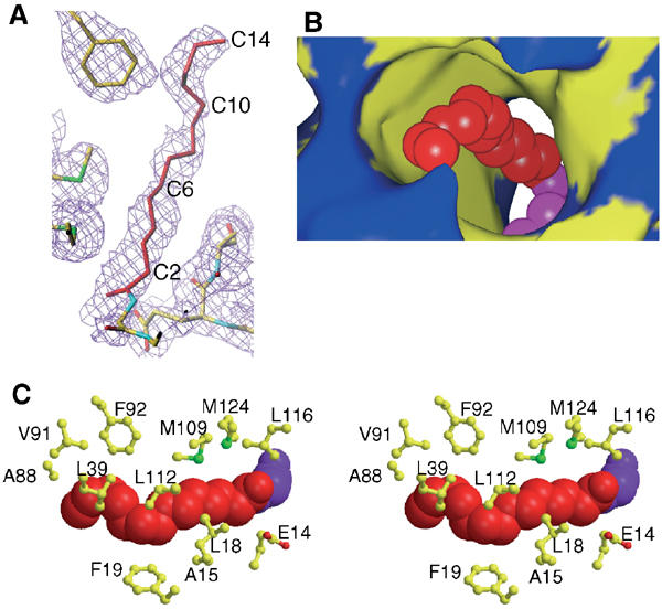
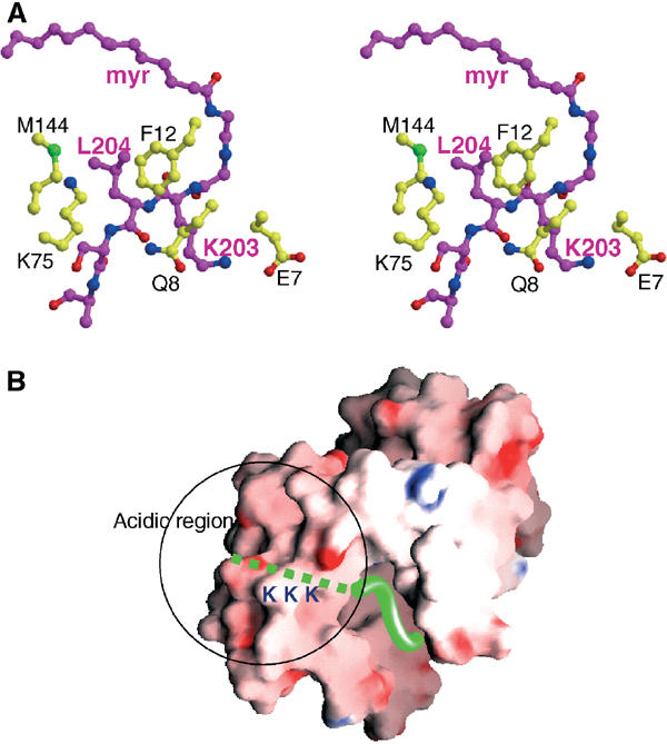
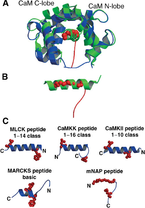
A variety of viral and signal transduction proteins are known to be myristoylated. Although the role of myristoylation in protein-lipid interaction is well established, the involvement of myristoylation in protein-protein interactions is less well understood. CAP-23/NAP-22 is a brain-specific protein kinase C substrate protein that is involved in axon regeneration. Although the protein lacks any canonical calmodulin (CaM)-binding domain, it binds CaM with high affinity. The binding of CAP-23/NAP-22 to CaM is myristoylation dependent and the N-terminal myristoyl group is directly involved in the protein-protein interaction. Here we show the crystal structure of Ca2+-CaM bound to a myristoylated peptide corresponding to the N-terminal domain of CAP-23/NAP-22. The myristoyl moiety of the peptide goes through a hydrophobic tunnel created by the hydrophobic pockets in the N- and C-terminal domains of CaM. In addition to the myristoyl group, several amino-acid residues in the peptide are important for CaM binding. This is a novel mode of binding and is very different from the mechanism of binding in other CaM-target complexes.


Figures

Figure 1
Figure 1
Crystal structure of Ca2+/CaM complexed with the mNAP peptide. Ribbon representation of the complex from the (A) front and (B) side. The CaM N- and C-terminal domains are shown in blue and green, respectively, the mNAP peptide is shown in red, and Ca2+ ions are shown as purple spheres. The figure was generated using MOLSCRIPT (Kraulis, 1991) and Raster3D (Merrit and Muphy, 1994). In the final model, Ala1, Asp2, Thr79, Asp80, Ala147 and Lys148 of CaM, and Lys207, Lys208 and Lys209 of the mNAP peptide are missing, while Glu11, Asp50, Lys77, Asp78, Lys105 and Glu127 of CaM, and Lys206 of the mNAP peptide, which have weak side chain densities, are modeled as alanines.
Figure 2
Figure 2
Binding interface of the myristoyl moiety of the mNAP peptide in the complex. (A) Electron density map of the myristoyl moiety. The simulated-annealing electron density map was refined by CNS (Brunger, 1998) and is contoured at 1.0σ. The myristoyl moiety is shown in red. The figure was generated using TURBO-FRODO (Roussel and Cambillau, 1991). (B) Hydrophobic tunnel of CaM–mNAP peptide complex. The space model of the myristoyl moiety is shown in red, and the hydrophobic residues of CaM are shown in yellow. The figure was prepared with SPOCK (Christopher, 1998). (C) Stereo view of CaM residues within 5 Å of the myristoyl group as generated by MOLSCRIPT (Kraulis, 1991) and Raster3D (Merrit and Muphy, 1994).
Figure 3
Figure 3
Binding interface of specific residues in CaM with the mNAP peptide. (A) Stereo representation of the binding mode in the N-terminal protein domain generated by MOLSCRIPT (Kraulis, 1991) and Raster3D (Merrit and Muphy, 1994). The myristoylated peptide is indicated in purple, and the residues of CaM are shown in yellow. (B) Electrostatic surface of CaM colored by charge (red, negative; blue, positive), with the mNAP peptide shown in ribbon (green) representation. The figure was produced using GRASP (Nicholls et al, 1991).
Figure 4
Figure 4
Comparison of the CaM–mNAP peptide with other CaM–peptide complexes. Images were generated by MOLSCRIPT (Kraulis, 1991) and Raster3D (Merrit and Muphy, 1994). (A) The CaM–mNAP peptide complex (blue) is superimposed onto the CaM–CaMKII peptide complex (green) to give the maximal alignment of the N-terminal domains. (B) Comparison of the space location of the bound peptides. The α-helix formed by the CaMKII peptide is shown in green, while the mNAP peptide is shown in red. (C) Comparison of CaM–peptide complexes. MLCK peptide (1–14 motif), CaMKK (1–16 motif), CaMKII (1–10 motif), MARCKS (basic motif) and mNAP peptide (novel motif) are shown. Side chains of the two key hydrophobic residues are shown in red.
Figure 5
Figure 5
(A) Interactions between the CaM C-terminal lobe and the bound peptides. The CaM–MLCK peptide (red, left side), the CaM–CaMKII peptide (green, left side) and the CaM–MARCKS peptide structure (yellow, left side) were superimposed onto the CaM–mNAP peptide structure (blue, right side) with respect to the C-terminal domain of CaM. The hydrophobic residues (Trp800 in MLCK, Leu299 in CaMKII and Phe157 in MARCKS) occupied the space in the hydrophobic pockets of the CaM C-terminal lobe. In contrast, the hydrophobic pocket was absent in CaM–mNAP peptide structure. (B) Interactions between the CaM N-terminal lobe and the bound peptides. The CaM–MLCK peptide (red, left side), the CaM–CaMKII peptide (green, left side) and the CaM–MARCKS peptide structure (yellow, left side) were superimposed onto the CaM–mNAP peptide structure (blue, right side) with respect to N-terminal domain of CaM. The hydrophobic residues (Leu813 in MLCK and Leu308 in CaMKII) occupied the space in the hydrophobic pockets of the CaM N-terminal lobe. In contrast, the hydrophobic pocket was absent in CaM–mNAP peptide structure.

Similar articles

Cited by

References

    1. Ames JB, Ishima R, Tanaka T, Gordon JI, Stryer L, Ikura M (1997) Molecular mechanics of calcium-myristoyl switches. Nature 389: 198–202 - PubMed
    1. Bomze HM, Bulsara KR, Iskandar BJ, Caroni P, Skene JHP (2001) Spinal axon regeneration evoked by replacing two growth cone proteins in adult neurons. Nat Neurosci 4: 38–43 - PubMed
    1. Brunger A, Adams PD, Clore GM, DeLano WL, Gros P, Grosse-Kunstleve RW, Jiang JS, Kuszewski J, Nilges M, Pannu NS, Read RJ, Rice LM, Simonson T, Warren GL (1998) Crystallography and NMR system: a new software suite for macromolecular structure determination. Acta Crystallogr D 54: 905–921 - PubMed
    1. Carr SA, Biemann K, Shoji S, Parmelee DC, Titani K (1982) n-Tetradecanoyl is the NH2-terminal blocking group of the catalytic subunit of cyclic AMP-dependent protein kinase from bovine cardiac muscle. Proc Natl Acad Sci USA 79: 6128–6131 - PMC - PubMed
    1. CCP4 (1994) The CCP4 suite: programs for protein crystallography. Acta Crystallogr D 50: 760–763 - PubMed

Publication types

Associated data