Structural basis of HP1/PXVXL motif peptide interactions and HP1 localisation to heterochromatin
- PMID: 14765118
- PMCID: PMC1271814
- DOI: 10.1038/sj.emboj.7600088
Structural basis of HP1/PXVXL motif peptide interactions and HP1 localisation to heterochromatin
Abstract
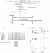
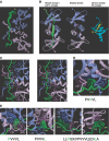
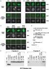
HP1 family proteins are adaptor molecules, containing two related chromo domains that are required for chromatin packaging and gene silencing. Here we present the structure of the chromo shadow domain from mouse HP1beta bound to a peptide containing a consensus PXVXL motif found in many HP1 binding partners. The shadow domain exhibits a novel mode of peptide recognition, where the peptide binds across the dimer interface, sandwiched in a beta-sheet between strands from each monomer. The structure allows us to predict which other shadow domains bind similar PXVXL motif-containing peptides and provides a framework for predicting the sequence specificity of the others. We show that targeting of HP1beta to heterochromatin requires shadow domain interactions with PXVXL-containing proteins in addition to chromo domain recognition of Lys-9-methylated histone H3. Interestingly, it also appears to require the simultaneous recognition of two Lys-9-methylated histone H3 molecules. This finding implies a further complexity to the histone code for regulation of chromatin structure and suggests how binding of HP1 family proteins may lead to its condensation.
Figures

Similar articles
-
The HP1 chromo shadow domain binds a consensus peptide pentamer.Curr Biol. 2000 Jan 13;10(1):27-30. doi: 10.1016/s0960-9822(99)00260-2. Curr Biol. 2000. PMID: 10660299
-
The structure of mouse HP1 suggests a unique mode of single peptide recognition by the shadow chromo domain dimer.EMBO J. 2000 Apr 3;19(7):1587-97. doi: 10.1093/emboj/19.7.1587. EMBO J. 2000. PMID: 10747027 Free PMC article.
-
Peptide recognition by heterochromatin protein 1 (HP1) chromoshadow domains revisited: Plasticity in the pseudosymmetric histone binding site of human HP1.J Biol Chem. 2017 Apr 7;292(14):5655-5664. doi: 10.1074/jbc.M116.768374. Epub 2017 Feb 21. J Biol Chem. 2017. PMID: 28223359 Free PMC article.
-
Biochemical and structural properties of heterochromatin protein 1: understanding its role in chromatin assembly.J Biochem. 2014 Jul;156(1):11-20. doi: 10.1093/jb/mvu032. Epub 2014 May 13. J Biochem. 2014. PMID: 24825911 Review.
-
How HP1 Post-Translational Modifications Regulate Heterochromatin Formation and Maintenance.Cells. 2020 Jun 12;9(6):1460. doi: 10.3390/cells9061460. Cells. 2020. PMID: 32545538 Free PMC article. Review.
Cited by
-
HP1α is a chromatin crosslinker that controls nuclear and mitotic chromosome mechanics.Elife. 2021 Jun 9;10:e63972. doi: 10.7554/eLife.63972. Elife. 2021. PMID: 34106828 Free PMC article.
-
HIRA vs. DAXX: the two axes shaping the histone H3.3 landscape.Exp Mol Med. 2024 Feb;56(2):251-263. doi: 10.1038/s12276-023-01145-3. Epub 2024 Feb 1. Exp Mol Med. 2024. PMID: 38297159 Free PMC article. Review.
-
Human heterochromatin proteins form large domains containing KRAB-ZNF genes.Genome Res. 2006 Dec;16(12):1493-504. doi: 10.1101/gr.5391806. Epub 2006 Oct 12. Genome Res. 2006. PMID: 17038565 Free PMC article.
-
H3K9me3-binding proteins are dispensable for SETDB1/H3K9me3-dependent retroviral silencing.Epigenetics Chromatin. 2011 Jul 20;4(1):12. doi: 10.1186/1756-8935-4-12. Epigenetics Chromatin. 2011. PMID: 21774827 Free PMC article.
-
The secret life of chromatin tethers.FEBS Lett. 2023 Nov;597(22):2782-2790. doi: 10.1002/1873-3468.14685. Epub 2023 Jun 25. FEBS Lett. 2023. PMID: 37339933 Free PMC article. Review.
References
-
- Bannister AJ, Zegerman P, Partridge JF, Miska EA, Thomas JO, Allshire RC, Kouzarides T (2001) Selective recognition of methylated lysine 9 on histone H3 by the HP1 chromo domain. Nature 410: 120–124 - PubMed
-
- Brown KE, Baxter J, Graf D, Merkenschlager M, Fisher AG (1999) Dynamic repositioning of genes in the nucleus of lymphocytes preparing for cell division. Mol Cell 3: 207–217 - PubMed
Publication types
MeSH terms
Substances
Associated data
- Actions
LinkOut - more resources
Full Text Sources
Molecular Biology Databases