. 2004 Feb 25;23(4):980-8.
doi: 10.1038/sj.emboj.7600086. Epub 2004 Feb 5.

An extracellular aspartic protease functions in Arabidopsis disease resistance signaling

Affiliations

An extracellular aspartic protease functions in Arabidopsis disease resistance signaling

Yiji Xia et al. EMBO J. .

Abstract

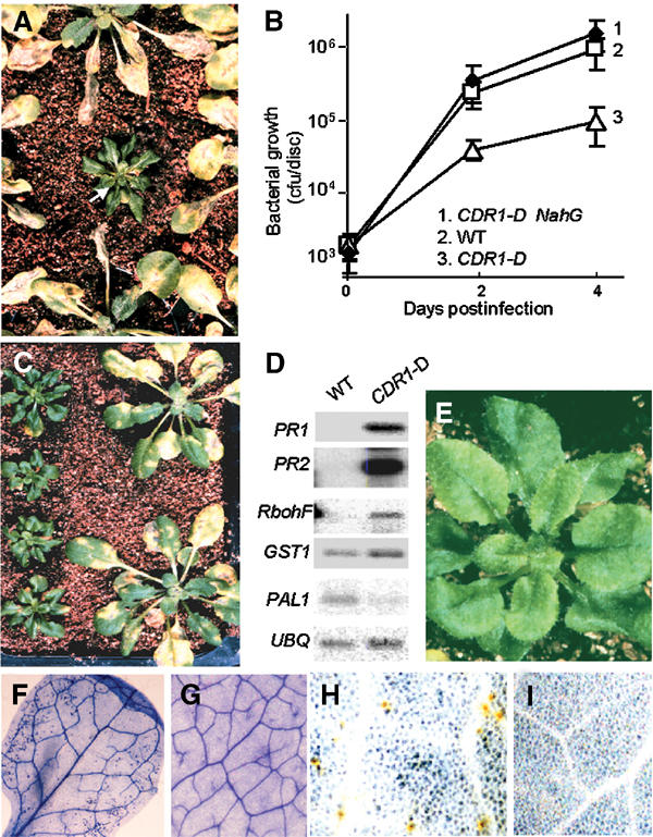
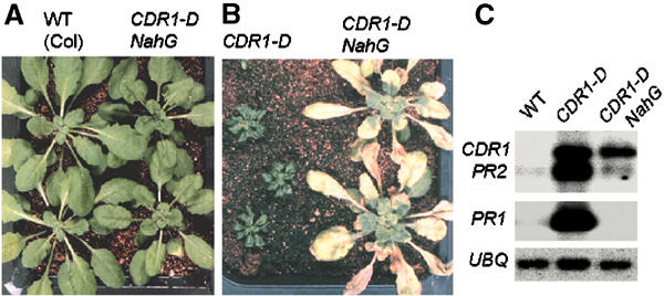
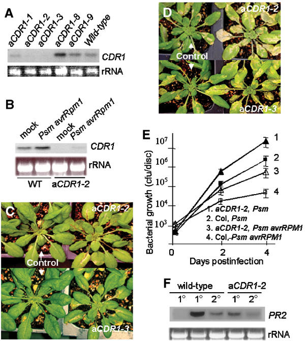
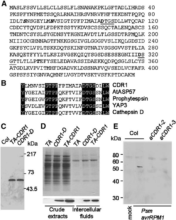
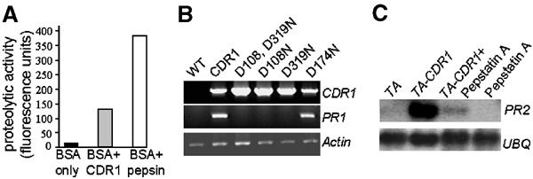
We have used activation tagging with T-DNA carrying cauliflower mosaic virus 35S enhancers to investigate the complex signaling networks underlying disease resistance in Arabidopsis. From a screen of approximately 5000 lines, we identified constitutive disease resistance (CDR1) encoding an apoplastic aspartic protease, the overexpression of which causes dwarfing and resistance to virulent Pseudomonas syringae. These phenotypes reflect salicylic-acid-dependent activation of micro-oxidative bursts and various defense-related genes. Antisense CDR1 plants were compromised for resistance to avirulent P. syringae and more susceptible to virulent strains than wild type. CDR1 accumulates in intercellular fluid in response to pathogen attacks. Induction of CDR1 generates a small mobile signal, and CDR1 action is blocked by the protease inhibitor pepstatin and by mutations in the protease active sites. We propose that CDR1 mediates a peptide signal system involved in the activation of inducible resistance mechanisms.


Figures

Figure 1
Figure 1
Visible and molecular phenotypes of the CDR1-D mutant. (A) Identification of the CDR1-D mutant in the T1 generation. T1 plants were infected by spraying with virulent Pst DC3000. (B) In planta growth of virulent Pst in CDR1-D, CDR1-D × NahG F1 plants and wild-type Col-0. Plants were infected by the dipping procedure. (C) CDR1-D exhibits enhanced resistance to Psm. CDR1-D T3 plants (left) and wild-type Col-0 plants (right) were infected by spraying with the virulent Psm strain M4. (D) Defense gene expression in uninfected CDR1-D plants compared to wild-type Col-0 plants. See text for details. UBQ is a ubiquitin gene used to control for RNA loading. (E) No obvious lesion formation is observed with the naked eye in CDR1-D. (F–I) Micro-oxidative bursts and micro-HRs in CDR1-D plants. (F) Trypan blue staining shows small clusters of dead cells throughout the leaf of an uninfected CDR1-D plant, but not in a wild-type plant (G). (H) DAB staining shows localized H2O2 production in uninfected CDR1-D plants, but not in control plants (I).
Figure 2
Figure 2
Involvement of SA in expression of CDR1-D phenotypes. (A) NahG suppresses the CDR1-D dwarf stature. (B) CDR1-D NahG plants no longer exhibit enhanced resistance to Pst. (C) CDR1-D NahG plants do not accumulate high levels of PR gene transcripts.
Figure 3
Figure 3
Cloning of CDR1. (A) Southern blot analysis showing the 10 kb EcoRI fragment that cosegregates with the CDR1-D mutant phenotype. (B) Structure of the CDR1-D allele. The rescued plasmid pCDR1E contains the 10 kb EcoRI fragment consisting of part of the T-DNA and the flanking 5 kb plant sequence. CDR1-like is likely a pseudogene that shares a sequence similarity (60% identify at the nucleotide level). (C) CDR1 expression is massively enhanced in the CDR1-D mutant. The bottom panel shows a portion of the RNA gel stained with ethidium bromide. (D) pBI/CDR1X construct containing the XbaI fragment of pCDR1E in the binary vector pBI101, which contains one copy of the 35S enhancer and the genomic fragment of CDR1. (E) Plants transformed with pBI/CDR1X exhibit the CDR1-D phenotypes of dwarf stature and resistance to Pst (wild-type Co-l plants are shown below). Only one of the transgenic lines was shown.
Figure 4
Figure 4
Phenotypes of the CDR1 antisense suppression lines. (A, B) Suppression of CDR1 expression in the three aCDR1 lines. RNA was isolated from uninfected wild type and the five aCDR1 lines (A) and from wild type and aCDR1-2 plants 8 h postinoculation with Psm avrRpm1 (B). Mock inoculation was used as a control. (C, D) aCDR1-2, aCDR1-3, and control plants containing an empty vector were infected by spraying with Psm avrRpm1 (C) and Psm (D). (E) In planta growth of Psm avrRpm1 and Psm in aCDR1-2 and control plants. (F) The antisense line is impaired in local and systemic induction of PR2 by inoculation of Psm avrRpm1. RNA was isolated from inoculated (1°, 8 h postinoculation) and systemic (2°, 48 h postinoculation) leaves of wild-type and the anti-CDR1 plants. The first lane is from mock-inoculated plants as a negative control.
Figure 5
Figure 5
CDR1 is an extracellular protein and is induced in response to bacterial inoculation. (A) Deduced amino acid sequence of CDR1. The two active sites are underlined, and the active site aspartyl residues are in bold. One potential N-glycosylation (N93) and two potential O-glycosylation sites (S85 and T329) are in italic. (B) Partial sequence alignment of CDR1 and four other aspartic proteases in the regions surrounding the first active site. (C) Affinity-purified anti-CDR1 polyclonal antibodies detect an approximately 54 kDa polypeptide in extracts from leaves of the CDR1-D mutant and from TA-CDR1 plants 16 h after Dex application. (D) Coomassie staining of total protein extracts and IFs from wild-type Col-0 plants carrying the empty vector (TA) and plants overexpressing CDR1 (upper panel) and immunodetection of CDR1 protein in the samples (lower panel). (E) CDR1 protein accumulates in IFs of wild-type plants but not the aCDR1 lines in response to bacterial inoculation. IFs were isolated from leaves 8 h after inoculation. The 54 kDa band represents CDR1, and other weak bands likely resulted from cross-reactions of the antibody to other proteins.
Figure 6
Figure 6
Induction of CDR1 leads to local and systemic activation of defense responses. (A) pTA-CDR1: construct for Dex inducible CDR1 expression. (B) Induction of TA-CDR1 by hand infiltration with Dex induces local and systemic PR gene expression. Two or three leaves of each plant were hand infiltrated with 5 μM Dex. The infiltrated (1°) and uninfiltrated (2°) leaves were collected separately. The TA plants carry the empty vector. (C) Grafting experiment showing induction of PR2 expression in wild-type scions grafted onto TA-CDR1 rootstocks following CDR1 induction. CDR1 was induced by watering with 10 μM Dex, and leaves were collected for RNA isolation 24 h after Dex application. Scions grafted onto TA rootstocks were used as a control. Lane 1: without Dex application; lanes 2 and 3: the samples from two sets of grafting experiments. (D) IFs isolated from Dex-treated TA-CDR1 plants induce PR gene expression. RNA was isolated from the wild-type leaves 10 h after hand infiltration with the IFs. IFs were isolated from TA transgenics (control) (16 h after spraying with 2 μM Dex, lane 1) and from TA-CDR1 transgenics either before (lane 3) or 16 h after (lane 2) Dex treatment. (E) Induction of PR2 by the IF was compromised in NahG and eds5-1 mutant. (F) Local and systemic induction of PR gene expression by HMW and LMW fractions of the IFs. (G) Immunodetection of CDR1 in the crude IF and two fractions of the IF. IF from TA transgenics was used as a negative control. (H) The elicitor in the LMW fraction has a molecular weight of 3–10 kDa. Only PR2 induction in the secondary leaves is shown. (I) The elicitor activity in the 3–10 kDa fraction is sensitive to heating and Pronase treatments.
Figure 7
Figure 7
CDR1 function requires aspartic protease activity. (A) In vitro proteolytic activity assay of E. coli-expressed CDR1. FITC-BSA was used as the substrate, and pepsin was used as a positive control. (B) RT–PCR analysis of PR gene expression in Arabidopsis transformed with CDR1 or various site-directed mutants in which active site or nonactive site aspartate residues are mutated. (C) Influence of pepstatin A on PR gene expression induced by IFs from CDR1 overexpressing Arabidopsis.

References

    1. Altschul SF, Gish W, Miller W, Myers EW, Lipman DJ (1990) Basic local alignment search tool. J Mol Biol 215: 403–410 - PubMed
    1. Alvarez ME, Pennell R, Meijer P-J, Ishikawa A, Dixon RA, Lamb C (1998) Reactive oxygen intermediates mediate a systemic signal network in the establishment of plant immunity. Cell 92: 773–784 - PubMed
    1. An G (1986) Development of plant promoter expression vectors and their use for analysis of differential activity of nopaline synthase promoter in transformed tobacco cells. Plant Physiol 81: 86–91 - PMC - PubMed
    1. Aoyama T, Chua N-H (1997) A glucocorticoid-mediated transcriptional induction system in transgenic plants. Plant J 11: 605–612 - PubMed
    1. Barrett AJ (1998) Proteolytic enzymes: nomenclature and classification. In Handbook of Proteolytic Enzymes, Woessner JF (ed) London: Academic Press pp 1–20

Publication types

MeSH terms

Substances

Associated data