. 2004 Feb 25;23(4):823-32.
doi: 10.1038/sj.emboj.7600079. Epub 2004 Feb 5.

DCX, a new mediator of the JNK pathway

Affiliations

DCX, a new mediator of the JNK pathway

Amos Gdalyahu et al. EMBO J. .

Abstract

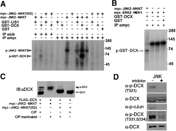
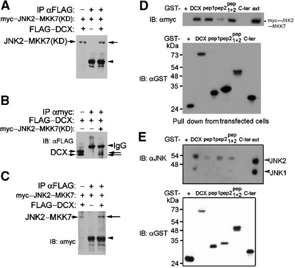
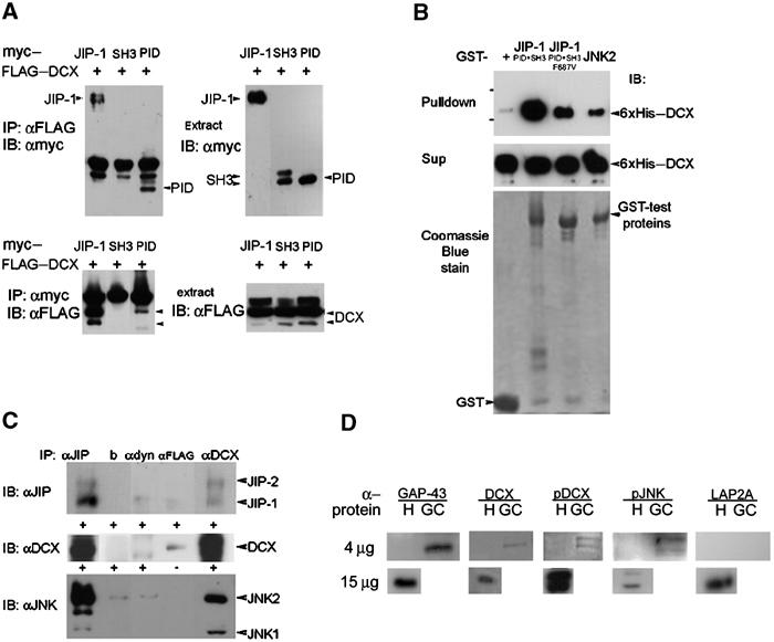
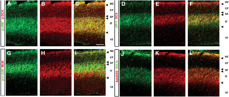
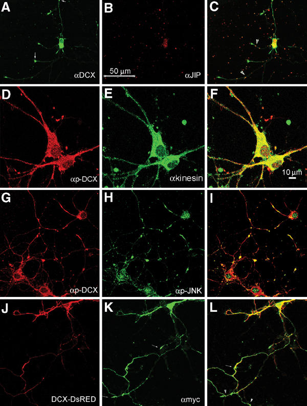
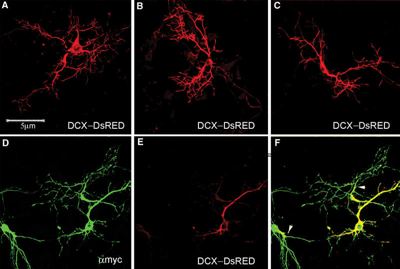
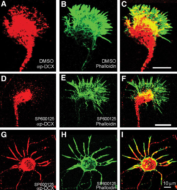
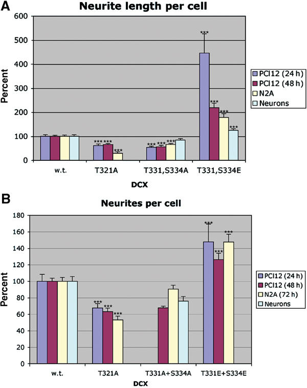
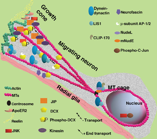
Mutations in the X-linked gene DCX result in lissencephaly in males, and abnormal neuronal positioning in females, suggesting a role for this gene product during neuronal migration. In spite of several known protein interactions, the involvement of DCX in a signaling pathway is still elusive. Here we demonstrate that DCX is a substrate of JNK and interacts with both c-Jun N-terminal kinase (JNK) and JNK interacting protein (JIP). The localization of this signaling module in the developing brain suggests its functionality in migrating neurons. The localization of DCX at neurite tips is determined by its interaction with JIP and by the interaction of the latter with kinesin. DCX is phosphorylated by JNK in growth cones. DCX mutated in sites phosphorylated by JNK affected neurite outgrowth, and the velocity and relative pause time of migrating neurons. We hypothesize that during neuronal migration, there is a need to regulate molecular motors that are working in the cell in opposite directions: kinesin (a plus-end directed molecular motor) versus dynein (a minus-end directed molecular motor).


Figures

Figure 1
Figure 1
DCX is a JNK substrate. (A) DCX is a substrate of JNK2–MKK7. Myc-tagged JNK2–MKK7 kinase active or dead (KD) were immunoprecipitated from transfected cells and used for in vitro phosphorylation assays using γ-ATP32, with the autoradiogram shown here. The kinase source was immunoprecipitated by anti-myc antibodies or anti-tubulin antibodies as a control. The recombinant proteins used in the assay were GST–DCX, GST–LIS1, and GST. (B) Erk2 does not phosphorylate DCX. ERK2–MKK1 was used in addition to JNK2–MKK7. Note that although the autophosphorylation of JNK2–MKK7 is less than that of ERK2–MKK1, GST–DCX is phosphorylated mainly by the first kinase and insignificantly by the second. (C) DCX is phosphorylated in vivo. Transfected DCX is phosphorylated in cells, since alkaline phosphatase (CIP) treatment reduced the mobility of DCX. In the presence of activated JNK, a significant mobility shift is noted that is reduced with CIP treatment. (D) DCX is phosphorylated in rat primary hippocampal neurons. Phosphorylation was detected in vivo by Western blot analysis using two sets of anti-p-DCX antibodies (designated T321, or T331, S334). Addition of the JNK inhibitor SP600125 resulted in abolishment of the signal of p-DCX as well as of p-cJun, although the amount of total proteins loaded was similar (see total DCX).
Figure 2
Figure 2
DCX and JNK interact. (AC) DCX and JNK co-immunoprecipitated from transfected cells. Cells were transfected with FLAG-tagged DCX, myc-tagged JNK2–MKK7 kinase dead or active as indicated. In each of the blots, the left lane is the extract, the middle lane is immunoprecipitations from cells transfected with one plasmid, and the right lane is the test immunoprecipitations. (A, C) Immunoprecipitations with anti-FLAG antibodies, immunoblot with anti-myc antibodies. (B) Immunoprecipitations with anti-myc antibodies, immunoblot with anti-FLAG antibodies. (D) Pull down experiments from transfected cells. Cells transfected with myc–JNK2–MKK7 kinase active were subject to GST pulldown using the following GST fusion proteins (order left to right): GST, DCX, pep1 (amino acids 51–135), pep2 (amino acids 178–259), pep1+2 (amino acids 51–259), Cter (from amino acid 273 to end). Note that both pep1 and pep2 interacted independently with myc–JNK2–MKK7. (E) Pull down experiments from P7 mouse brain extracts. The following were the recombinant proteins (order left to right): GST, GST–DCX, GST–pep1, GST–pep2, GST–pep1+2, GST–Cter. The GST proteins were checked by immunoblot (lower blots in D, E).
Figure 3
Figure 3
DCX interacts with JIP-1 and the interaction domain is in the DC motif and the PID domain, respectively. (A) Cells transfected with FLAG–DCX, and with myc-tagged JIP-1, the SH3 domain of JIP-1, or the PID domain of JIP-1 were subjected to immunoprecipitations using anti-FLAG antibodies (top left panel) or anti-myc antibodies (bottom left panel). The expression of each of the proteins was verified by Western blot analysis (both right panels). The interaction domain mapped within the PID domain. (B) Recombinant proteins 6xHis-tagged DCX were pulled down using the corresponding GST-tagged proteins (GST control, JIP-1 (SH3+PID domain), JIP-1 (SH3+PID domain mutated F687V), JNK2). The levels of the input proteins were similarly judged by Coomassie blue stain (lower panel) or anti-His Western blot (middle panel). (C) DCX, JIP and JNK co-immunoprecipitated from mouse brain extracts. P7 mouse brain was used for immunoprecipitations using monoclonal anti-DCX antibodies (228), anti-FLAG antibodies (Sigma), anti-dynein antibodies (Sigma), beads, or anti-JIP goat polyclonal antibodies (E19, Santa Cruz), followed by Western blot analysis using anti-JIP1 mouse monoclonal antibodies (BD Transduction Laboratories), anti-JNK2 mouse monoclonal antibodies (D-2, Santa Cruz), and anti-DCX rabbit polyclonal antibodies (Shmueli et al, 2001). The negative controls were anti-FLAG, anti-dynein, and beads only. DCX antibodies immunoprecipitated JIP-1 and JIP-2 (top panel), JIP-1 antibodies immunoprecipitated DCX (middle panel), and DCX antibodies immunoprecipitated JNK1 and JNK2 (lower panel). (D) DCX, p-DCX, and p-JNK are enriched in growth cones. Homogenates (H) (4 or 15 μg) and growth-cone preparations (GC) (4 μg) were separated on gels and Western blotted with the following antibodies: GAP-43 (positive control, enriched in growth cones), DCX, p-DCX, p-JNK (these proteins are also enriched in growth cones), and LAP2A (negative control, nuclear protein).
Figure 4
Figure 4
Localization of the JNK module in the developing cerebral cortex. (AL) Coronal sections of E15.5 cortex stained with antibodies against components of the JNK module: pJNK (Thr 183, Tyr 185) p-DCX (A–C) DCX, JIP1 (D–F), MUK the MAPKKK and a substrate of JNK, phosphorylated c-JUN (G–I), ApoER2, reelin (Reln) (JL). Images represent one optical slice (2.5 μm).
Figure 5
Figure 5
Localization of DCX and the JNK signaling module molecules in rat primary hippocampal neurons. (AC) Colocalization of DCX and JIP-1 in some primary hippocampal neurons. DCX stained in green; some of the tips are marked with arrowheads (A), JIP in red (B) and some of the overlapping dots are indicated (arrowheads) (C). (DF) Colocalization of p-DCX (D) and conventional kinesin (E), overlap in (F). (GI) Colocalization of p-DCX (G) and p-JNK (H), merge in (I), in primary hippocampal neurons pretreated with kainic acid. (JL) DCX–DsRED and JIP-1 colocalized in transfected neurons: DCX–DsRED (J) myc-tagged-JIP-1 stained with anti-myc antibodies (in green, K), merge (L), note the labeled tips of the neurites (small arrows, L).
Figure 6
Figure 6
Expression of dominant-negative JIP-1 results in mislocalization of DCX–DsRED. (AC) DCX–DsRED is well distributed in transfected primary hippocampal neurons. (DF) Cotransfection of DCX–DsRED with myc–JIP-1 dominant negative. myc–JIP-1 dominant negative is well distributed in the neurons (in green, D), and DCX–DsRED remains less distributed (in red, E) as indicated in the overlap (F, arrowheads).
Figure 7
Figure 7
p-DCX intracellular localization. (A) Rat primary hippocampal growth cone stained with anti-p-DCX, T331, S334 (red) and (B) phalloidin-FITC (green); note the high degree of overlap (C) (yellow). (DI) Growth cones treated with a specific JNK inhibitor and then stained with anti-p-DCX, T331, S334 (D), or T321 (G) (red) and phalloidin-FITC (green, E, H); note the reduced overlap (F, I, in comparison to C).
Figure 8
Figure 8
Effect of unphospho- and phospho-mimicking DCX mutants on neuronal cells. PC12, N2A, and primary cerebellar cells were transfected with the indicated DCX mutant constructs, and cells were analyzed for neurite length using image J (A) and number of neurites per cell (B). The s.e. is indicated in the bars. ***P<0.001.
Figure 9
Figure 9
Model of a neuron migrating along radial glia. This model is based on earlier models (Morris et al, 1998; Feng and Walsh, 2001; Gupta et al, 2002; Hatten, 2002) and incorporates the finding and hypotheses derived from this paper. The migrating neuron has an elongated structure with a growth cone. There is more p-DCX located in the growth cone where it interacts with JNK and JIP. JIP is mobilized there by kinesin. JIP also interacts with ApoER2 that binds to the extracellular matrix protein reelin, and to kinesin that is a plus-end directed motor. DCX also interacts with the membranal protein neurofascin, and with the μ-subunits of the AP-1/2 complexes. At the MT plus-end tips we can also find CLIP-170, which recruits LIS1, and the dynein–dynactin complex. The dynein–dynactin retrograde motor is recruited to MTs with LIS1 and DCX followed by enhanced activity of this motor. Nucleokinesis is assisted by the activity of the dynein motor that is associated with the MT cage and the centrosome (there also mNudE and NudeL can be found). Within the nucleus, the transcription factor c-Jun is phosphorylated by JNK. The activity of JNK may thereby indirectly regulate the differential activities of kinesin and dynein.

References

    1. Aicardi J (1989) The lissencephaly syndromes. Int Pediat 4: 118–126
    1. Assadi AH, Zhang G, Beffert U, McNeil RS, Renfro AL, Niu S, Quattrocchi CC, Antalffy BA, Sheldon M, Armstrong DD, Wynshaw-Boris A, Herz J, D'Arcangelo G, Clark GD (2003) Interaction of reelin signaling and Lis1 in brain development. Nat Genet 35: 270–276 - PubMed
    1. Barth PG (1987) Disorders of neuronal migration. Can J Neurol Sci 14: 1–16 - PubMed
    1. Bennett BL, Sasaki DT, Murray BW, O'Leary EC, Sakata ST, Xu W, Leisten JC, Motiwala A, Pierce S, Satoh Y, Bhagwat SS, Manning AM, Anderson DW (2001) SP600125, an anthrapyrazolone inhibitor of Jun N-terminal kinase. Proc Natl Acad Sci USA 98: 13681–13686 - PMC - PubMed
    1. Biernat J, Mandelkow EM (1999) The development of cell processes induced by tau protein requires phosphorylation of serine 262 and 356 in the repeat domain and is inhibited by phosphorylation in the proline-rich domains. Mol Biol Cell 10: 727–740 - PMC - PubMed

Publication types

MeSH terms