. 2004 Feb 11;23(3):605-15.
doi: 10.1038/sj.emboj.7600074. Epub 2004 Feb 5.

A Suv39h-dependent mechanism for silencing S-phase genes in differentiating but not in cycling cells

Affiliations

A Suv39h-dependent mechanism for silencing S-phase genes in differentiating but not in cycling cells

Slimane Ait-Si-Ali et al. EMBO J. .

Abstract

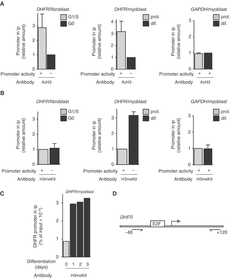
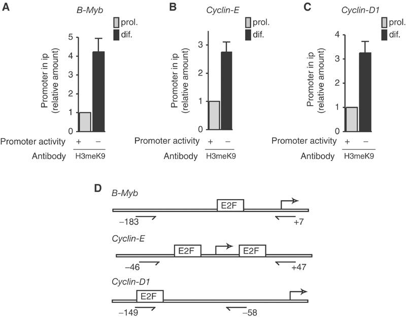
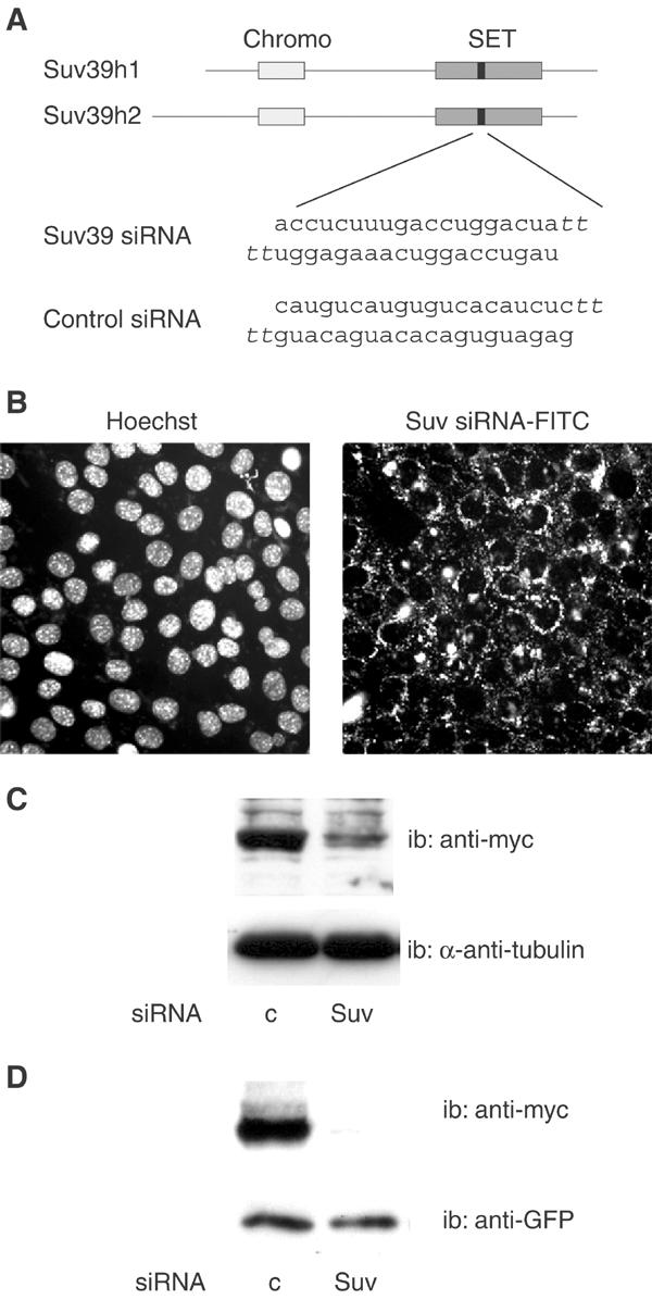
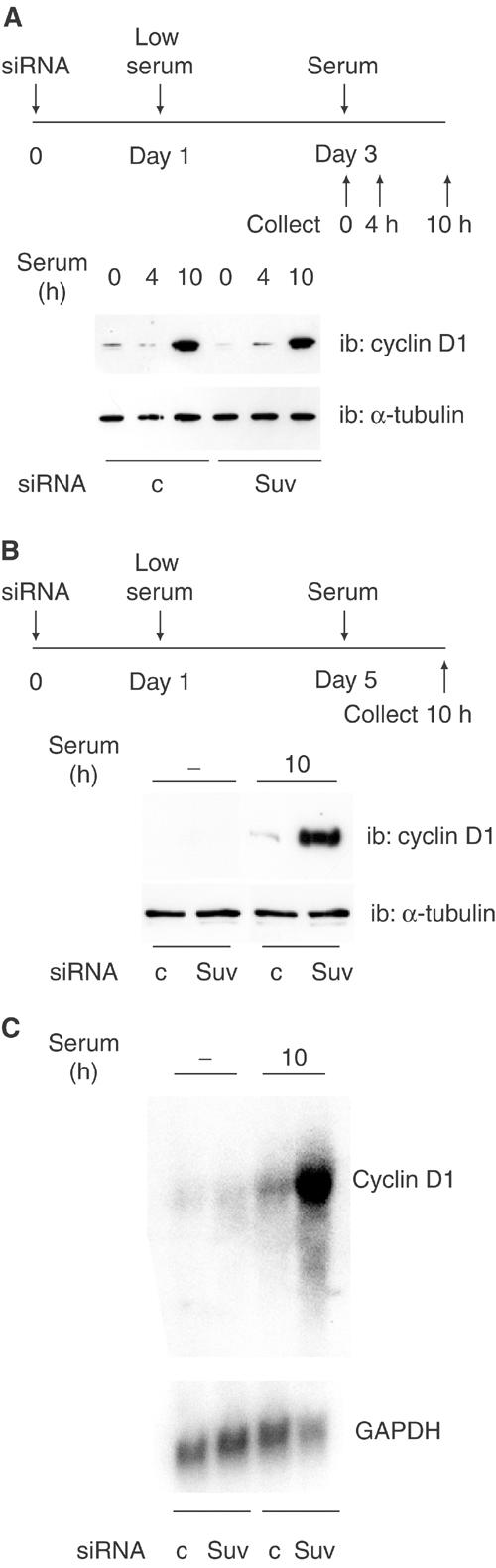
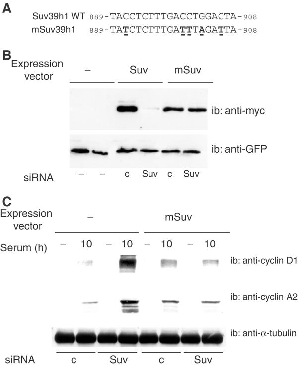
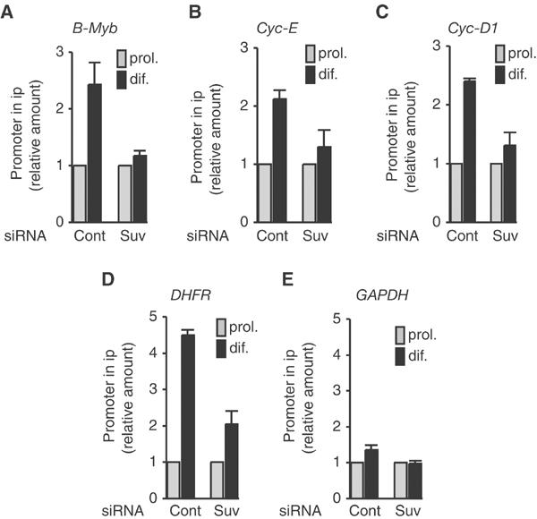
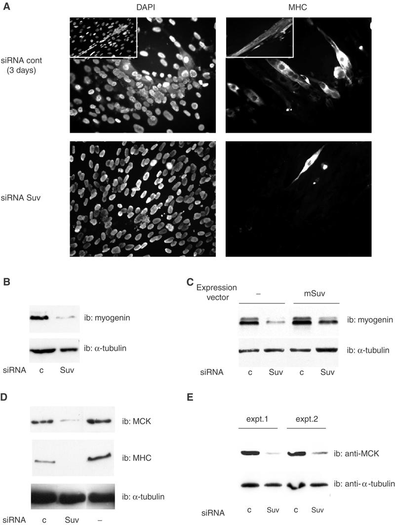
The Rb/E2F complex represses S-phase genes both in cycling cells and in cells that have permanently exited from the cell cycle and entered a terminal differentiation pathway. Here we show that S-phase gene repression, which involves histone-modifying enzymes, occurs through distinct mechanisms in these two situations. We used chromatin immunoprecipitation to show that methylation of histone H3 lysine 9 (H3K9) occurs at several Rb/E2F target promoters in differentiating cells but not in cycling cells. Furthermore, phenotypic knock-down experiments using siRNAs showed that the histone methyltransferase Suv39h is required for histone H3K9 methylation and subsequent repression of S-phase gene promoters in differentiating cells, but not in cycling cells. These results indicate that the E2F target gene permanent silencing mechanism that is triggered upon terminal differentiation is distinct from the transient repression mechanism in cycling cells. Finally, Suv39h-depleted myoblasts were unable to express early or late muscle differentiation markers. Thus, appropriately timed H3K9 methylation by Suv39h seems to be part of the control switch for exiting the cell cycle and entering differentiation.


Figures

Figure 1
Figure 1
Histone modifications at the DHFR promoter in fibroblasts or myoblasts as indicated. Chromatin was prepared from NIH3T3 fibroblasts at different stages of the cell cycle (G0 or G1/S as indicated, see Materials and methods) or from C2C12 myoblastic cells, either proliferating (prol.) or after either 2 days (dif.) (A, B) or indicated period of time (C) in differentiation medium. Chromatin was immunoprecipitated with antibodies directed against pan-acetylated H3 (AcH3 (A)), or methylated K9 histone H3 (H3meK9 (B, C)) as indicated, and analyzed by Q-PCR to quantify the DHFR promoter copy number, or the GAPDH gene (negative control) copy number. H3 acetylation results (mean±s.d., n=3) are shown as the ratio between values from cells in G1/S and G0 phase (for fibroblasts) or as the ratio of data for proliferating versus differentiating cells (for myoblasts). H3K9 methylation results (mean±s.d., n=3) are shown as the ratio between values from cells in G0 and G1/S phase (for fibroblasts), as the ratio of data for differentiating versus proliferating cells (for myoblasts), or as the percent of input chromatin immunoprecipitated using anti-H3meK9 antibodies (C). Promoter activity was assessed by Northern blot (see Ferreira et al, 2001). (D) Schematic representation of the DHFR promoter showing the positions of the E2F site, transcription start site (bent arrow) and primers used for PCR (with reference to the transcription start site).
Figure 2
Figure 2
Methylation of H3K9 at various S-phase gene promoters. ChIP experiments were performed as described in Figure 1 and assayed for B-Myb (A), Cyclin-E (B) and Cyclin-D1 (C) promoters (mean±s.d., n=4). (D) Schematic representations of the promoters and positions of the primers as in Figure 1.
Figure 3
Figure 3
Expression of Suv39h1 mRNA in muscle cells. RNA from C2C12 cells either proliferating (0 h) or after different periods of differentiation were analyzed by Q-RT–PCR. The results are shown after standardization on GAPDH mRNAs quantified in the same experiments.
Figure 4
Figure 4
Inhibition of Suv39h expression by siRNAs. (A) Sequence of Suv39h siRNA and position of its target in the mRNAs (black boxes) corresponding to positions 889–908 for Suv39h1 and 1111–1130 for Suv39h2 (chromo: chromodomain; SET: SET domain). The scrambled sequence used as a control is also shown (Control siRNA). SiRNAs contain two 3′ overhanging T's. (B) Penetration of Suv39h siRNA into myoblast. Proliferating C2C12 cells were transfected with FITC-labeled Suv39h siRNA, and examined 18 h later. (C) Inhibition of Suv39h1 protein in HeLa cells. A HeLa-derived human cell line stably expressing a myc-tagged version of Suv39h1 (see Materials and methods) was transfected with Suv39h (Suv) or control (c) siRNAs, and analyzed 40 h later by Western blotting using anti-myc antibodies, or anti-α-tubulin antibodies as a control. (D) Inhibition of Suv39h protein in myoblasts. C2C12 cells were cotransfected with Suv39 siRNA or control siRNA, together with the myc-tagged Suv39h1 expression vector and a GFP expression vector used as a transfection control. At 48 h post-transfection, cell lysates were analyzed by Western blotting with anti-myc or anti-GFP.
Figure 5
Figure 5
Suv39h inhibition affects differentiating cells but not cycling cells. (A) NIH3T3 cells were treated with control siRNA or Suv39h siRNA, synchronized and treated with serum as described in the upper panel; cyclin D1 expression was monitored by Western blotting. Suv39h1 mRNA was downregulated by 84% in the experiment shown. (B) C2C12 cells were treated with control siRNA or Suv39h siRNA, placed in differentiation medium and treated with serum as indicated in the upper panel, and analyzed as in (A). Suv39h1 mRNA was downregulated by 86% in the experiment shown. (C) C2C12 cells were treated with control siRNA or Suv39h siRNA as in (B), and RNA was extracted and analyzed by Northern blot using indicated probes. Inhibition of Suv39h was 79% as measured by RT–PCR. These experiments were repeated 2–4 times.
Figure 6
Figure 6
Rescue experiments using a Suv39h siRNA-resistant mutant. (A) Sequences of wild type and mutant Suv39h; conservative mutations are underlined. (B) C2C12 cells were cotransfected with Suv39 siRNA or control siRNA, together with the indicated expression vectors: myc-tagged Suv39h1 wild type (Suv), myc-tagged Suv39h siRNA-resistant mutant (mSuv) or the empty vehicle vector (−); a GFP expression vector was used as a transfection control. At 48 h post-transfection, cell lysates were analyzed by Western blotting with anti-myc or anti-GFP. (C) C2C12 cells were cotransfected with Suv39h siRNA (Suv) or control siRNA (c) together with either the Suv39h siRNA-resistant mutant (mSuv) expression vector or the empty vehicle vector (−), and treated as in Figure 5B; extracts were analyzed by Western blotting using anti-cyclin D1 or -cyclin A2 antibodies, as indicated. Suv39h1 mRNA was downregulated by 80% in the experiment shown. These experiments were repeated twice.
Figure 7
Figure 7
Suv39h depletion affects H3K9 methylation at S-phase gene promoters. C2C12 cells were transfected with control siRNA (cont) or Suv39h siRNA (Suv) as indicated. At 48 h post-transfection, cells were placed in differentiation medium for 72 h and chromatin was prepared and immunoprecipitated as in Figure 1. S-phase gene promoters were detected in immunoprecipitates as described in Figure 2 (mean±s.d., n=3–4).
Figure 8
Figure 8
Suv39h depletion affects the muscle differentiation program. (A, B) C2C12 cells were transfected with control siRNA or Suv39h siRNA as indicated. At 48 h post-transfection, cells were placed in differentiation medium for 72 h (or 96 h for control siRNA-transfected cells in the insets; no mature myotubes were observed in Suv39 siRNA-transfected cells under the same conditions) and labeled by immunofluorescence using an anti-MHC antibody (A) or analyzed by Western blotting with anti-myogenin (B). (C) C2C12 cells were transfected as in (A), together with the expression vector for the siRNA-resistant form of Suv39h (mSuv) or the empty vehicle vector for the control; differentiation was induced 24 h after transfection. (D) C2C12 cells were transfected as in (A), and extracts were analyzed by Western blotting with anti-MHC and -MCK antibodies. Suv39h mRNA was downregulated by 80–86% in these experiments. These experiments were repeated 2–3 times. (E) Primary myoblasts were transfected with siRNAs using Lipofectamine (experiment 1) or Lipofectamine 2000 (experiment 2); inhibition of Suv39h was 74.1 and 73.9%, respectively. Cells were placed in differentiation medium for 2 days, and assayed for MCK expression.

References

    1. Aagaard L, Laible G, Selenko P, Schmid M, Dorn R, Schotta G, Kuhfittig S, Wolf A, Lebersorger A, Singh PB, Reuter G, Jenuwein T (1999) Functional mammalian homologues of the Drosophila PEV-modifier Su(var)3-9 encode centromere-associated proteins which complex with the heterochromatin component M31. EMBO J 18: 1923–1938 - PMC - PubMed
    1. Aagaard L, Schmid M, Warburton P, Jenuwein T (2000) Mitotic phosphorylation of SUV39H1, a novel component of active centromeres, coincides with transient accumulation at mammalian centromeres. J Cell Sci 113: 817–829 - PubMed
    1. Bannister AJ, Zegerman P, Partridge JF, Miska EA, Thomas JO, Allshire RC, Kouzarides T (2001) Selective recognition of methylated lysine 9 on histone H3 by the HP1 chromo domain. Nature 410: 120–124 - PubMed
    1. Brehm A, Miska EA, McCance DJ, Reid JL, Bannister AJ, Kouzarides T (1998) Retinoblastoma protein recruits histone deacetylase to repress transcription. Nature 391: 597–601 - PubMed
    1. Buckingham M (1996) Skeletal muscle development and the role of the myogenic regulatory factors. Biochem Soc Trans 24: 506–509 - PubMed

Publication types

MeSH terms