. 2004 Feb 25;23(4):739-48.
doi: 10.1038/sj.emboj.7600066. Epub 2004 Feb 5.

The phagocyte NADPH oxidase depends on cholesterol-enriched membrane microdomains for assembly

Affiliations

The phagocyte NADPH oxidase depends on cholesterol-enriched membrane microdomains for assembly

Frederik Vilhardt et al. EMBO J. .

Abstract

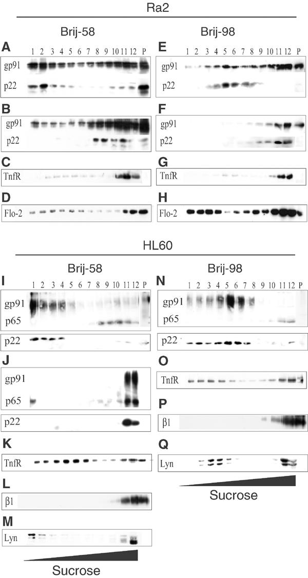
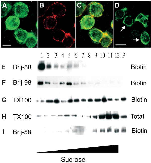
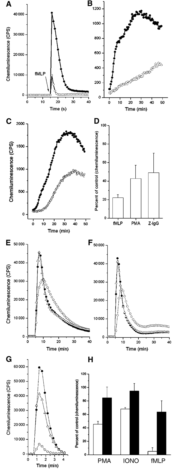
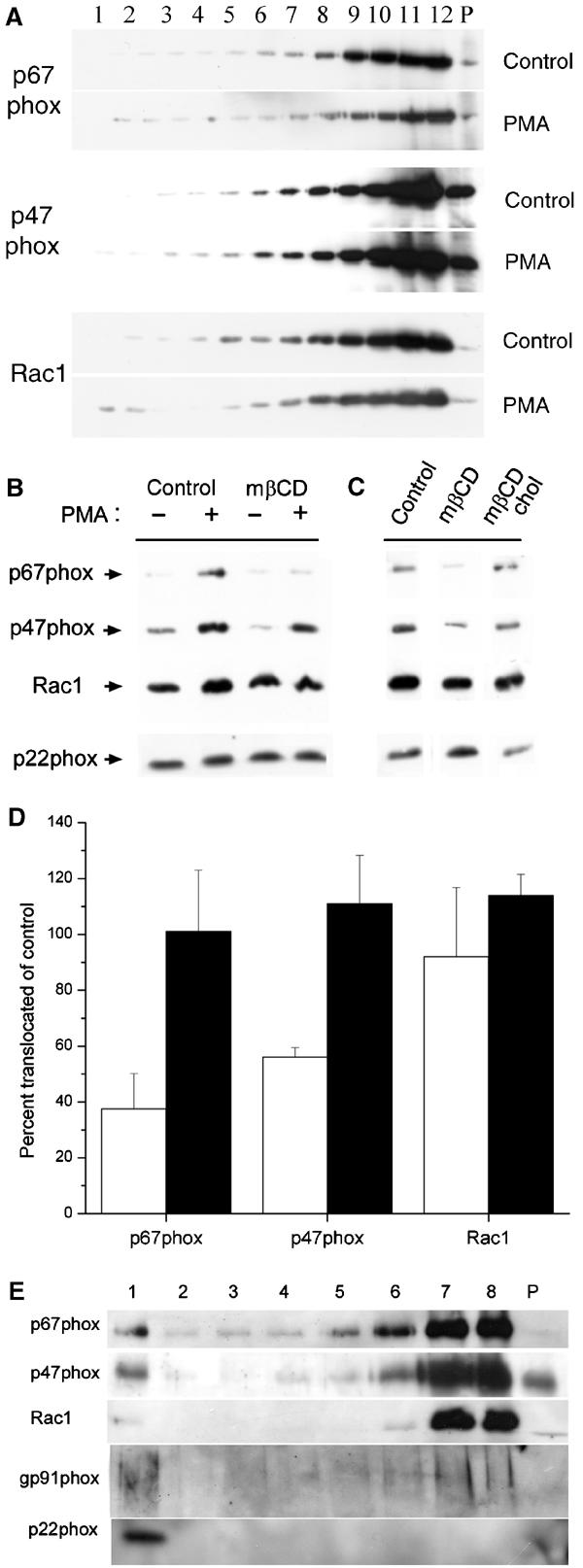
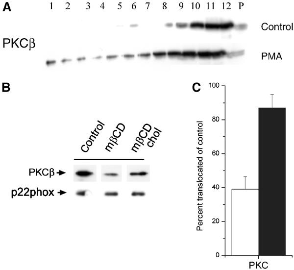
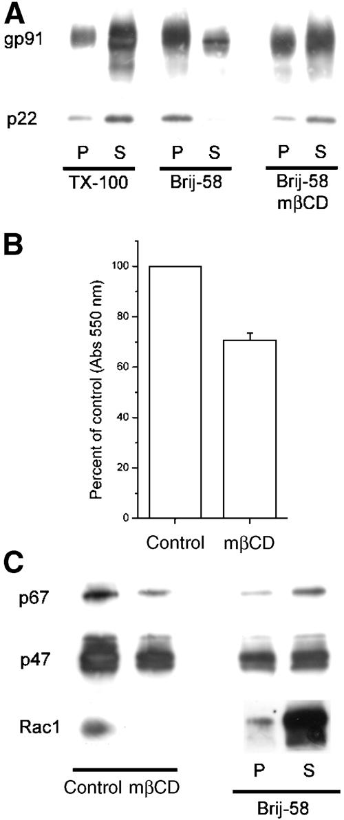
The superoxide-producing phagocyte NADPH oxidase consists of a membrane-bound flavocytochrome b558 complex, and cytosolic factors p47phox, p67phox and the small GTPase Rac, which translocate to the membrane to assemble the active complex following cell activation. We here show that insolubility of NADPH oxidase subunits in nonionic detergents TX-100, Brij-58, and Brij-98 is a consequence of inclusion into cholesterol-enriched membrane microdomains (lipid rafts). Thus, flavocytochrome b558, in a cholesterol-dependent manner, segregated to the bouyant low-density detergent-resistant membrane (DRM) fraction, and the cytosolic NADPH oxidase factors associated dynamically with low-density DRM. Further, superoxide production following cholesterol depletion was severely compromised in intact cells or in a cell-free reconstituted system, correlating with a reduced translocation of cytosolic phox subunits to the membrane. In analogy with the widely accepted role of lipid rafts as signaling platforms, our data indicate that cholesterol-enriched microdomains act to recruit and/or organize the cytosolic NADPH oxidase factors in the assembly of the active NADPH oxidase.


Figures

Figure 1
Figure 1
Gp91phox and p22phox segregate to the buoyant low-density detergent (Brij-58, Brij-98) resistant membrane fraction. Ra2 microglia (A–H) or HL60 (I–Q) cells were lysed in Brij-58 (A–D, I–M) or Brij-98 (E–H, N–Q), in some cases with prior mβCD-facilitated cholesterol extraction (B, F, J), and the lysate was centrifuged to equilibrium in a 35–15% sucrose gradient. Recovered fractions (no. 12 is the bottom, high-density fraction of the gradient) and the solubilized pellet (P) were analyzed by Western blotting using antibodies against gp91phox (gp91), p22phox (p22), transferrin receptor (TfnR), β1-integrin (β1), flotillin-2 (Flo-2), or Lyn as indicated.
Figure 2
Figure 2
Flavocytochrome b558 association with DRM is dependent on the subcellular localization of the complex. (A–C) Ra2 cells were incubated on ice with a ricin-HRP conjugate to label the cell surface before fixation. Subsequently, (A) gp91phox was localized with polyclonal anti-gp91phox antibodies, and (B) the cell surface visualized with anti-HRP monoclonal antibody. Panel (C) shows the merged channels, and illustrates that a substantial fraction of gp91phox is present intracellularly. (D) For comparison, gp91phox in differentiated HL60 cells is almost exclusively confined to the plasma membrane (arrows). Bars (A–D), 10 μm. (EI) Ra2 cells were surface-biotinylated and extracted in (E, I) Brij-58, (F) Brij-98 or (G, H) TX-100. Subsequent to sucrose gradient centrifugation, biotinylated protein, or in the case of TX-100, also total protein, was analyzed by Western blotting with anti-gp91phox antibodies (E–H) or anti-TnfR antibodies (I).
Figure 3
Figure 3
Gp91phox cosegregates with patched ganglioside GM1. GM1 or TnfR on HL60 cells was labeled with Alexa 488-conjugated cholera toxin B subunit (D–F) or anti-transferrin receptor antibodies (G–I), respectively, and subsequently patched by the addition of secondary antibodies (D–I). Subsequently, immunolocalization of gp91phox (B, E, H) was performed. (A–C) shows control cells stained with Alexa 488-conjugated cholera toxin B subunit without secondary antibody patching. Arrows point to areas of colocalization of gp91phox and GM1, while arrowheads denote areas with no apparent overlap. Note the pronounced copatching of gp91phox with GM1 (F), but not transferrin receptor (I). Bars 10 μm.
Figure 4
Figure 4
Cholesterol extraction disrupts compartmentalization of NADPH oxidase activity on the plasma membrane. Control (A, C) or cholesterol-extracted (B, D) Ra2 microglia cells were stained with filipin (A, B) to illustrate the efficiency of cholesterol removal. Asterisks denote intracellular stores of cholesterol (which are partially obscured in (A) due to the intense plasma membrane staining of control cells). Arrows point to plasma membrane staining, which is hardly visible in (B). (C, D) Cells were stimulated with PMA in the presence of NBT. Note the pronounced patchy distribution of the precipitation product in the control cells (C), and that cholesterol extraction causes the deposit to become diffusely distributed on the plasma membrane (D). Framed boxes are shown at 2 × magnification. Bars (A–D), 10 μm.
Figure 5
Figure 5
NADPH oxidase activity in Ra2 microglia and HL60 cells requires cholesterol. (A–D) Ra2 control (•) or cholesterol-extracted cells (○) were stimulated with (A) fMLP, (B) PMA, or (C) IgG-opsonized zymosan particles, and superoxide production measured continuously by luminol-enhanced chemiluminescence. Bar graph (D) shows mean and s.d. of three independent experiments carried out as in (A–C). Superoxide production of control cells was assigned an arbitrary value of 100. (E–H) Control (•), mβCD-extracted (○), or mβCD-extracted and then cholesterol-reconstituted (▵) HL60 cells were stimulated with either (E) PMA, (F) ionomycin, or (G) fMLP. Subsequently, superoxide production was measured as above. Bar graph (H) shows mean and s.d. of at least three independent experiments, expressed as percent superoxide production of control cells. Empty bars represent cholesterol-extracted cells, and filled bars represent cholesterol-replenished cells.
Figure 6
Figure 6
P47phox and p67phox segregation to low-density detergent (Brij-58) resistant membrane is increased by cell activation, and their membrane translocation is cholesterol-dependent. (A) Brij-58 lysates of control or PMA-stimulated HL60 cells were centrifuged to equilibrium in a sucrose gradient, and then analyzed by Western blotting using antibodies to p67phox, p47phox, or Rac1. (B) Control or cholesterol-extracted HL60 cells were stimulated with PMA, and subsequently the particulate membrane fraction was analyzed for the presence of p67phox, p47phox, or Rac1 by Western blotting. Equal aliquots were also analyzed for p22phox as loading control. (C) HL60 cells were subjected to cholesterol extraction (mβCD), in some cases followed by cholesterol replenishment (mβCD chol), before stimulation with PMA. p67phox, p47phox, Rac1, and p22phox was analyzed by Western blotting as described above. (D) The graph represents mean and s.e. of three independent experiments performed as in (C). Individual p67phox, p47phox, and Rac1 bands were quantitated densitometrically, and the results expressed as percent translocation in cholesterol-extracted (white bars) or cholesterol-replenished (filled bars) cells relative to control cells. (E) HL60 cells stimulated with PMA were subjected to chemical crosslinking with DTSP and the particulate membrane fraction, purified as above, was subjected to sucrose gradient centrifugation. Collected fractions were electrophoresed under reducing conditions and analyzed by Western blotting with anti-p67phox, p47phox, Rac1, gp91phox, and p22phox antibodies.
Figure 7
Figure 7
PKC membrane translocation is inhibited after mβCD-mediated cholesterol extraction. (A) Brij-58 lysates of control or PMA-stimulated HL60 cells were centrifuged to equilibrium in a sucrose gradient, and then analyzed by Western blotting using antibodies to PKCβ. (B) HL60 cells were subjected to cholesterol extraction (mβCD), in some cases followed by cholesterol replenishment (mβCD chol), before stimulation with PMA, and translocation of PKCβ to the particulate membrane fraction analyzed by Western blotting. Equal aliquots were also analyzed for p22phox as loading control. (C) The graph represents mean and s.e. of three independent experiments performed as in (B). The results are expressed as percent translocation in cholesterol-extracted (empty bars) or cholesterol-replenished (filled bars) cells relative to control cells.
Figure 8
Figure 8
NADPH oxidase assembly and activity in a reconstituted cell-free system depends on cholesterol. (A) Control or mβCD-treated purified macrophage membranes were extracted in TX-100 or Brij-58, and distribution of cyt b558 subunits in the detergent-insoluble pellet (P) and soluble fraction (S) was determined by Western blotting. (B) Superoxide production of control or mβCD-extracted purified macrophage membranes in the reconstituted amphiphile-activated system as measured by cytochrome c reduction. Results are expressed as percent cytochrome c reduction of control, and represent mean and s.d. of four independent experiments. Superoxide production of control membranes corresponded to 15.9 mol superoxide/mol cyt b558 heme/s. (C) Control or mβCD-extracted membranes mixed with cytosolic subunits were pelleted, and association of p67phox, p47phox, and Rac1 with membrane determined by lysis in Laemmli buffer and Western blotting. (D) Distribution of cytosolic subunits in detergent-insoluble pellet (P) and soluble fraction (S) after extraction of control membranes in Brij-58.

Similar articles

Cited by

References

    1. Ago T, Kuribayashi F, Hiroaki H, Takeya R, Ito T, Kohda D, Sumimoto H (2003) Phosphorylation of p47phox directs phox homology domain from SH3 domain toward phosphoinositides, leading to phagocyte NADPH oxidase activation. Proc Natl Acad Sci USA 100: 4474–4479 - PMC - PubMed
    1. Ago T, Nunoi H, Ito T, Sumimoto H (1999) Mechanism for phosphorylation-induced activation of the phagocyte NADPH oxidase protein p47(phox). Triple replacement of serines 303, 304, and 328 with aspartates disrupts the SH3 domain-mediated intramolecular interaction in p47(phox), thereby activating the oxidase. J Biol Chem 274: 33644–33653 - PubMed
    1. Allen LA, DeLeo FR, Gallois A, Toyoshima S, Suzuki K, Nauseef WM (1999) Transient association of the nicotinamide adenine dinucleotide phosphate oxidase subunits p47phox and p67phox with phagosomes in neutrophils from patients with X-linked chronic granulomatous disease. Blood 93: 3521–3530 - PubMed
    1. Bodin S, Giuriato S, Ragab J, Humbel BM, Viala C, Vieu C, Chap H, Payrastre B (2001) Production of phosphatidylinositol 3,4,5-trisphosphate and phosphatidic acid in platelet rafts: evidence for a critical role of cholesterol-enriched domains in human platelet activation. Biochemistry 40: 15290–15299 - PubMed
    1. Bokoch GM, Bohl BP, Chuang TH (1994) Guanine nucleotide exchange regulates membrane translocation of Rac/Rho GTP-binding proteins. J Biol Chem 269: 31674–31679 - PubMed

Publication types