

Isolation and characterization of a novel single-stranded RNA virus infecting the bloom-forming diatom Rhizosolenia setigera
- PMID: 14766545
- PMCID: PMC348932
- DOI: 10.1128/AEM.70.2.704-711.2004
Isolation and characterization of a novel single-stranded RNA virus infecting the bloom-forming diatom Rhizosolenia setigera
Abstract
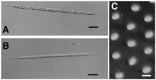
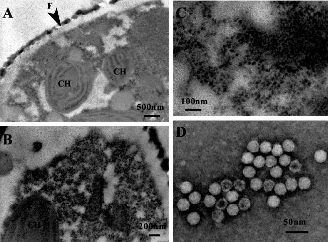
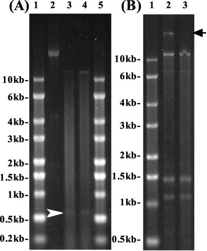
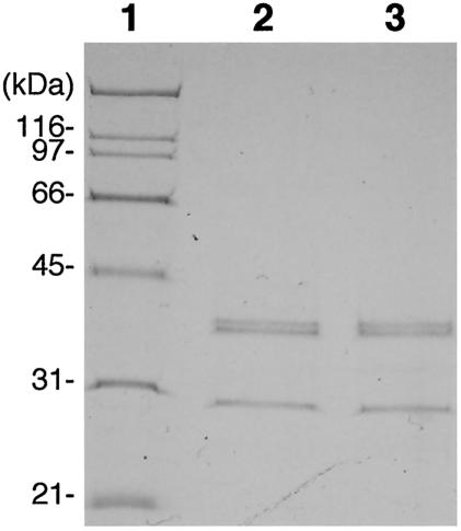
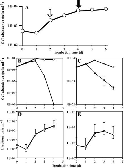
A novel single-stranded RNA (ssRNA) virus specifically infecting the bloom-forming diatom Rhizosolenia setigera (R. setigera RNA virus [RsRNAV]) was isolated from Ariake Sea, Japan. Viral replication occurred within the cytoplasm, and the virus particle was icosahedral, lacked a tail, and was 32 nm in diameter on average. The major nucleic acid extracted from the RsRNAV particles was an ssRNA molecule 11.2 kb in length, although smaller RNA molecules (0.6, 1.2, and 1.5 kb) were occasionally observed. The major structural proteins of RsRNAV were 41.5, 41.0, and 29.5 kDa. Inter- and intraspecies host specificity tests revealed that RsRNAV is not only species specific but also strain specific and that its intraspecies host specificity is diverse among virus clones. The latent period of RsRNAV was 2 days, and the burst sizes were 3,100 and 1,010 viruses per host cell when viruses were inoculated into the host culture at the exponential and stationary growth phases, respectively, at 15 degrees C under a 12-h-12-h light-dark cycle of ca. 110 micro mol of photons m(-2) s(-1) with cool white fluorescent illumination. To our knowledge, this is the first report describing the biological properties of a virus infecting a diatom. Further studies on RsRNAV will be helpful in understanding the ecological relationship between diatoms and viruses in nature.
Figures
References
-
- Bratbak, G., J. K. Egge, and M. Heldal. 1993. Viral mortality of the marine alga Emiliania huxleyi (Haptophyceae) and termination of algal blooms. Mar. Ecol. Prog. Ser. 93:39-48.
-
- Brussaard, C. P. D., R. S. Kempers, A. J. Kop, R. Riegman, and M. Heldal. 1996. Virus-like particles in a summer bloom of Emiliania huxleyi in the North Sea. Aquat. Microb. Ecol. 10:105-113.
-
- Chen, L. C. M., T. Edelstein, and J. McLachlan. 1969. Bonnemaisonia hamifera Hariot in nature and in culture. J. Phycol. 5:211-220. - PubMed
-
- Culley, A. I., A. S. Lang, and C. A. Suttle. 2003. High diversity of unknown picorna-like viruses in the sea. Nature 424:1054-1057. - PubMed
Publication types
MeSH terms
Substances
LinkOut - more resources
Full Text Sources
