. 2004 Feb;70(2):1040-50.
doi: 10.1128/AEM.70.2.1040-1050.2004.

Biochemical and proteomic analysis of the magnetosome membrane in Magnetospirillum gryphiswaldense

Affiliations

Biochemical and proteomic analysis of the magnetosome membrane in Magnetospirillum gryphiswaldense

Karen Grünberg et al. Appl Environ Microbiol. 2004 Feb.

Abstract

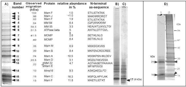
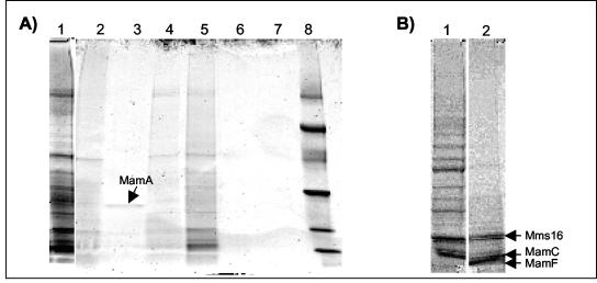
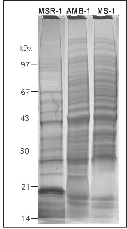
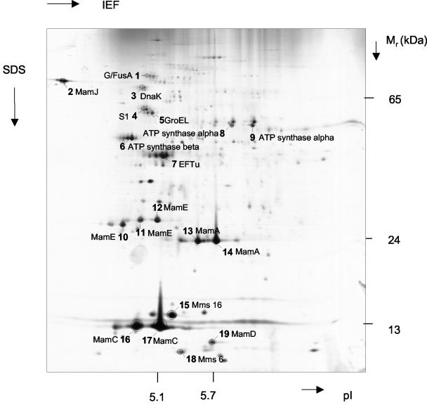
We analyzed the biochemical composition of the magnetosome membrane (MM) in Magnetospirillum gryphiswaldense. Isolated magnetosomes were associated with phospholipids and fatty acids which were similar to phospholipids and fatty acids from other subcellular compartments (i.e., outer and cytoplasmic membranes) but were present in different proportions. The binding characteristics of MM-associated proteins were studied by selective solubilization and limited proteolysis. The MM-associated proteins were further analyzed by various proteomic approaches, including one- and two-dimensional sodium dodecyl sulfate-polyacrylamide gel electrophoresis followed by Edman and mass spectrometric (electrospray ionization-mass spectrometry-mass spectrometry) sequencing, as well as capillary liquid chromatography-mass spectrometry-mass spectrometry of total tryptic digests of the MM. At least 18 proteins were found to constitute the magnetosome subproteome, and most of these proteins are novel for M. gryphiswaldense. Except for MM22 and Mms16, all bona fide MM proteins (MMPs) were encoded by open reading frames in the mamAB, mamDC, and mms6 clusters in the previously identified putative magnetosome island. Eight of the MMPs display homology to known families, and some of them occur in the MM in multiple homologues. Ten of the MMPs have no known homologues in nonmagnetic organisms and thus represent novel, magnetotactic bacterium-specific protein families. Several MMPs display repetitive or highly acidic sequence patterns, which are known from other biomineralizing systems and thus may have relevance for magnetite formation.


Figures

FIG. 1.
FIG. 1.
Separation of MM-associated proteins by 1D PAGE. (A) Summary of MMPs detected by Coomassie blue staining in 1D SDS-16% PAGE gels. (B and C) Coomassie blue-stained (B) and silver-stained 16% SDS-Tricine gels of MMPs. The heterogeneous band at 9 kDa (band 17) revealed an ambiguous N-terminal amino acid sequence, M(G/Y/F/I)(P/Q/T/K)(L/I/V)(K/A)(M/G/V)(A/T/V/I), which partially contains the N terminus of MamG. (D) Heme staining of MMPs (lane 1), which were subsequently stained with Coomassie blue (lane 2). The arrowheads indicate the positions of heme-positive bands. aa, amino acid.
FIG. 2.
FIG. 2.
Binding characteristics of the MM-associated proteins. (A) SDS-PAGE of proteins solubilized from the MM after treatment with various agents. Lane 1, 1% hot SDS (5 min); lane 2, Triton X-100 (2%); lane 3, Tween 20 (2%); lane 4, octylglucoside (500 mM); lane 5, SDS (5%); lane 6, urea (5 M); lane 7, NaCl (2 M); lane 8, low-molecular-weight marker. The arrow indicates the position of a 24-kDa band which was selectively solubilized by Tween 20 and was identified as MamA by its N-terminal amino acid sequence (MSSKPSN). (B) Lane 1, proteins from untreated magnetosomes; lane 2, proteins from magnetosomes after tryptic digestion. The arrows indicate the positions of protein bands resistant to partial tryptic digestion, which were identified as Mms16 (ASKQAEQLFD), MamC (MSFQLAPYLAK), and MamF (MAETILIETKT).
FIG. 3.
FIG. 3.
Electron micrographs of isolated magnetosome particles. (A) Untreated magnetosomes; (B) magnetosomes after treatment with Triton X-100; (C) magnetosomes after tryptic digestion; (D) magnetosomes after boiling with 1% SDS.
FIG. 4.
FIG. 4.
SDS-16% PAGE of MMPs from M. gryphiswaldense MSR-1, Magnetospirillum strain AMB-1, and M. magnetotacticum MS-1.
FIG. 5.
FIG. 5.
Silver-stained 2D PAGE of MMPs from M. gryphiswaldense. Proteins from marked spots were excised from the corresponding Coomassie blue-stained gel and identified by electrospray ionization-MS-MS after tryptic digestion. IEF, isoelectric focusing.
FIG. 6.
FIG. 6.
Selected sequence characteristics of several MMPs. (A) Sequence alignment of MamG and MamD of M. gryphiswaldense. (B) Dot blot analysis of MamJ by Pustell protein matrix analysis (39) (pam 250 matrix).
FIG. 7.
FIG. 7.
Molecular organization of a region from the putative magnetosome island of M. gryphiswaldense comprising the entire mms6, mamDC, and mamAB gene clusters (45). The solid arrows indicate genes encoding MMPs, which were identified in this study.

References

    1. Altschul, S. F., T. L. Madden, A. A. Schaffer, J. Zhang, Z. Zhang, W. Miller, and D. J. Lipman. 1997. Gapped BLAST and PSI-BLAST: a new generation of protein database search programs. Nucleic Acids Res. 25:3389-3402. - PMC - PubMed
    1. Appel, R. D., P. M. Palagi, D. Walther, J. R. Vargas, J. C. Sanchez, F. Ravier, C. Pasquali, and D. F. Hochstrasser. 1997. Melanie II-A third-generation software package for analysis of two-dimensional electrophoresis images. I. Features and user interface. Electrophoresis 18:2724-2734. - PubMed
    1. Arakaki, A., J. Webb, and T. Matsunaga. 2003. A novel protein tightly bound to bacterial magnetic particles in Magnetospirillum magneticum strain AMB-1. J. Biol. Chem. 278:8745-8750. - PubMed
    1. Baeuerlein, E. 2000. Single magnetic crystals of magnetite (Fe3O4) synthesized in intracytoplasmic vesicles of Magnetospirillum gryphiswaldense, p. 61-80. In E. Baeuerlein (ed.), Biomineralization. Wiley-VCH, Weinheim, Germany.
    1. Balkwill, D., D. Maratea, and R. Blakemore. 1980. Ultrastructure of a magnetotactic spirillum. J. Bacteriol. 141:1399-1408. - PMC - PubMed

Publication types

MeSH terms

Associated data

LinkOut - more resources