. 2004 Feb 17;101(7):1981-6.
doi: 10.1073/pnas.0307317101. Epub 2004 Feb 6.

Aberrant expansion of segmented filamentous bacteria in IgA-deficient gut

Affiliations

Aberrant expansion of segmented filamentous bacteria in IgA-deficient gut

Keiichiro Suzuki et al. Proc Natl Acad Sci U S A. .

Abstract

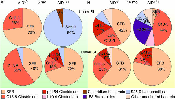
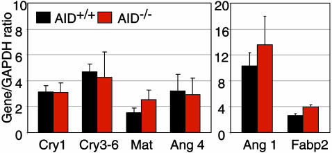
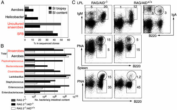
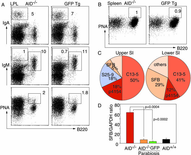
The mechanism to maintain homeostasis of the gut microbiota remains largely unknown despite its critical role in the body defense. In the intestines of mice with deficiency of activation-induced cytidine deaminase (AID), the absence of hypermutated IgA is partially compensated for by the presence of large amounts of unmutated IgM and normal expression levels of defensins and angiogenins. We show here a predominant and persistent expansion of segmented filamentous bacteria throughout the small intestine of AID(-/-) mice. Reconstitution of lamina propria IgA production in AID(-/-) mice recovered the normal composition of gut flora and abolished the local and systemic activation of the immune system. The results indicate that secretions of IgAs rather than innate defense peptides are critical to regulation of commensal bacterial flora and that the segmented filamentous bacteria antigens are strong stimuli of the mucosal immune system.


Figures

Fig. 1.
Fig. 1.
SFB expansion in AID–/– small intestine. Shown are intestinal flora in the biopsies from upper and lower segments of the small intestine of 5-month-old (A) and 16-month-old (B) AID–/– and AID+/+ mice. Biopsies were pooled from two littermates for each genotype, and the microflora was identified by sequence analyses of the 16S rRNA PCR products. Each color represents a specific sequence, and numbers indicate percentages in sequenced clones.
Fig. 2.
Fig. 2.
Innate immune defenses are not perturbed by commensal anaerobe expansion. Data are cryptdin, matrilysin, and angiogenin 4 mRNA expression by using RT-qPCR in the IEC preparations from small intestines of 4- to 5-month-old AID–/– and AID+/+ mice (n = 3, duplicate determinations; mean gene/GAPDH ratio ± SD plotted). Expression of angiogenin 1 and fatty acid binding protein (Fabp2) are showed as controls for IEC.
Fig. 3.
Fig. 3.
Anaerobe expansion in RAG-2–/– mice is reversed by AID+/+ but not AID–/– BM transplantation. (A) Small intestinal microflora in biopsy samples (black bars) and intestinal content (gray bars) of RAG-2–/– mice identified by using bacterial 16S rRNA universal primers. Samples were from 10-week-old mice (n = 4). (B) Microflora identified by microbiological methods in the small intestinal contents of RAG-2–/– mice (gray bars), RAG-2–/– mice injected with BM from AID+/+ mice (white bars), or RAG-2–/– mice injected with BM from AID–/– mice (black bars). (C) Fluorescence-activated cell sorting profiles of LP and spleen stained for B220, IgM, IgA, and peanut agglutinin (PNA) from the RAG-2–/– mice transferred with BM from AID–/– or AID+/+ mice. Numbers indicate the lymphocyte percentages in the gates. Data showed in B and C are from the same animals and are representative profiles from two sets of mice.
Fig. 4.
Fig. 4.
IgA reconstitution in AID–/– mice rescue the gut microbiota and B cell activation phenotype. (A and B) Representative fluorescence-activated cell sorting profiles of LPL and spleen stained for B220, IgM, IgA, and peanut agglutinin (PNA) from AID–/– and GFP Tg mice 6 months after anastomosis. Numbers indicate the percentages of lymphocytes in the gates. (C) Intestinal flora in the biopsies from upper and lower segments of the small intestine of AID–/– mice after parabiosis with GFP Tg mice. Results shown in AC are from the same animals and are representative profiles from four sets of mice. (D) Quantitative PCR for SFB in IEC preparations from AID–/– mice (red bar), AID+/+ mice (black bar), and AID–/– mice after parabiosis with GFP Tg mice (orange and green bars, respectively). Data are mean ± SE; n = 4; P values (unpaired Student t test) are shown.

References

    1. Cebra, J. J. (1999) Am. J. Clin. Nutr. 69, 1046S–1051S. - PubMed
    1. Hooper, L. V. & Gordon, J. I. (2001) Science 292, 1115–1118. - PubMed
    1. Ayabe, T., Satchell, D. P., Wilson, C. L., Parks, W. C., Selsted, M. E. & Ouellette, A. J. (2000) Nat. Immunol. 1, 113–118. - PubMed
    1. Hooper, L. V., Stappenbeck, T. S., Hong, C. V. & Gordon, J. I. (2003) Nat. Immunol. 4, 269–273. - PubMed
    1. Salzman, N. H., Ghosh, D., Huttner, K. M., Paterson, Y. & Bevins, C. L. (2003) Nature 422, 522–526. - PubMed

Publication types

MeSH terms

Substances