. 2004 Apr;15(4):1904-17.
doi: 10.1091/mbc.e04-01-0004. Epub 2004 Feb 6.

The PXL1 gene of Saccharomyces cerevisiae encodes a paxillin-like protein functioning in polarized cell growth

Affiliations

The PXL1 gene of Saccharomyces cerevisiae encodes a paxillin-like protein functioning in polarized cell growth

Nancy A Mackin et al. Mol Biol Cell. 2004 Apr.

Abstract

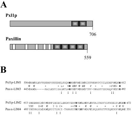
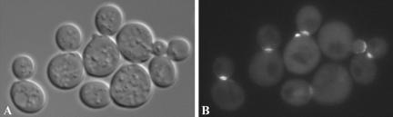
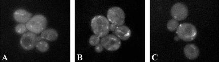
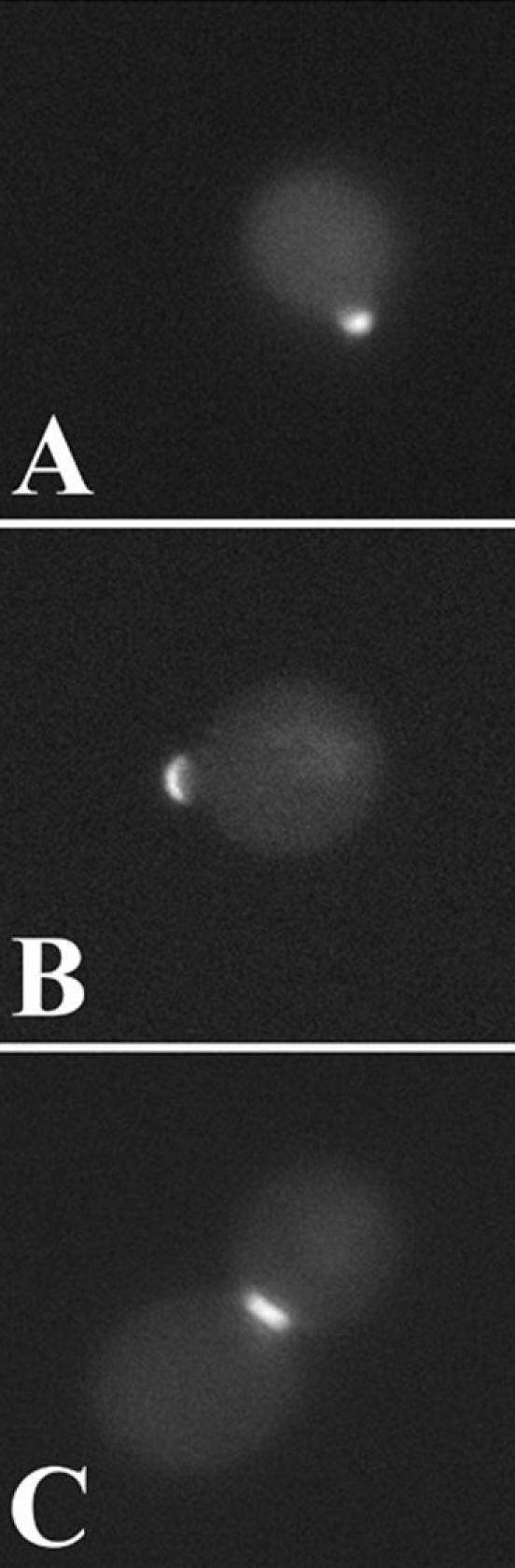
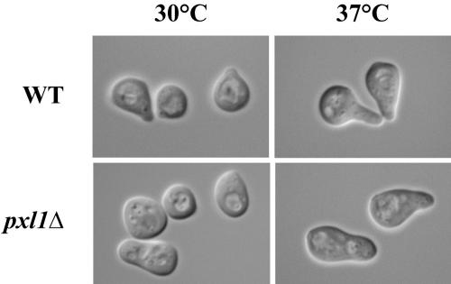
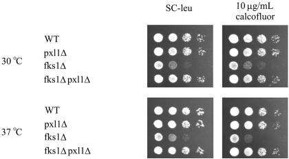
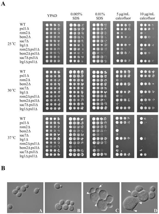
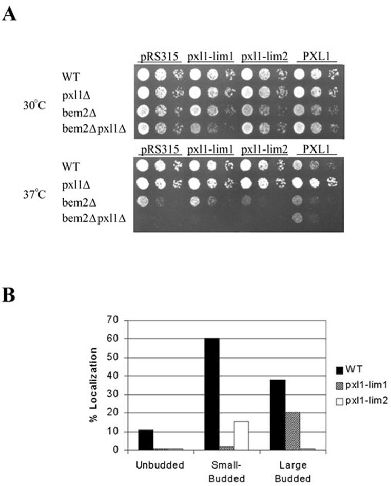
The Saccharomyces cerevisiae open reading frame YKR090w encodes a predicted protein displaying similarity in organization to paxillin, a scaffolding protein that organizes signaling and actin cytoskeletal regulating activities in many higher eucaryotic cell types. We found that YKR090w functions in a manner analogous to paxillin as a mediator of polarized cell growth; thus, we have named this gene PXL1 (Paxillin-like protein 1). Analyses of pxl1Delta strains show that PXL1 is required for the selection and maintenance of polarized growth sites during vegetative growth and mating. Genetic analyses of strains lacking both PXL1 and the Rho GAP BEM2 demonstrate that such cells display pronounced growth defects in response to different conditions causing Rho1 pathway activation. PXL1 also displays genetic interactions with the Rho1 effector FKS1. Pxl1p may therefore function as a modulator of Rho-GTPase signaling. A GFP::Pxl1 fusion protein localizes to sites of polarized cell growth. Experiments mapping the localization determinants of Pxl1p demonstrate the existence of localization mechanisms conserved between paxillin and Pxl1p and indicate an evolutionarily ancient and conserved role for LIM domain proteins in acting to modulate cell signaling and cytoskeletal organization during polarized growth.


Figures

Figure 1.
Figure 1.
(A) Schematics representing the structural organization of Pxl1p and paxillin. Shaded and solid boxes indicate LIM domains and LD repeats, respectively. Predicted protein lengths are shown. (B) Alignment of Pxl1p LIM domains and the LIM domains three and four of paxillin alpha. Numbers indicate amino acid residues. Boldface cysteine and histidine residues indicate the LIM domain consensus sequence. Symbols show amino acid identities (:) and similarities (+) between Pxl1p and paxillin family members.
Figure 2.
Figure 2.
Pxl1p localizes to sites of polarized growth. A MATa pxl1Δ strain (YSE554) expressing a GFP::Pxl1p fusion was examined using differential interference contrast microscopy (A) and epifluorescence microscopy (B). The localization of GFP::Pxl1p to polarized growth site locations can be seen to be cell cycle stage dependent.
Figure 3.
Figure 3.
The preLIM domain of Pxl1p localizes to punctate intracellular domains. (A and B) Wild-type and (C) pxl1Δ cells carrying the plasmid GFP::pxl1-1-502, encompassing the pre-LIM domain of Pxl1p but lacking the C-terminal LIM domains, were examined by epifluorescence microscopy for localization of the fusion protein. The fusion protein is associated with several punctate patches that lie within the interior of the cells.
Figure 4.
Figure 4.
The LIM domain containing C-terminus of Pxl1p is sufficient to target the protein to sites of polarized growth. (A-C) pxl1Δ cells carrying the plasmid GFP::pxl1-503-706 that lacks the preLIM domain of Pxl1p were examined at different stages of the cell cycle for the distribution of the fusion protein. GFP::pxl1-503-706 localizes to the incipient bud site (A), the growing bud tip (B), and the mother-bud neck (C), in a manner identical to the localization pattern of the full-length GFP::Pxl1p fusion protein.
Figure 5.
Figure 5.
Haploid pxl1Δ mutants possess defects in mating projection formation and morphology. At 30°, pxl1Δ cells form blunt mating projections compared with the narrow, focused projection tips characteristic of wild-type cells. Similarly, at 37° wild-type cells form extended or hyperpolarized projections in contrast to the blunt projections formed by pxl1Δ cells.
Figure 6.
Figure 6.
Removing PXL1 function suppresses the growth defect of cells lacking FKS1. Wild-type, pxl1Δ, fks1Δ, and pxl1Δ fks1Δ strains were examined for their growth rates on SC-leu medium containing 10 μg/ml calcofluor. Deletion of PXL1 can be seen to increase the growth rate of the pxl1Δ fks1Δ relative to the fks1Δ strain.
Figure 7.
Figure 7.
PXL1 is a specific genetic interactor with BEM2. (A) Growth of wild-type (YSE430), pxl1Δ (YSE554), rom2Δ (YSE637), bem2Δ (YSE661), sac7Δ (YSE818), and lrg1Δ (YSE804) strains was compared with double mutants lacking both PXL1 and one of the RHO1 GEF or GAP regulators: pxl1Δ rom2Δ (YSE665), pxl1Δ bem2Δ (YSE838), pxl1Δ sac7Δ (YSE814), and pxl1Δ lrg1Δ (YSE816). Growth of the indicated strains was examined at 25, 30, and 37°C on YPAD medium or YPAD supplemented with the cell wall destabilizing agents calcofluor white or SDS as indicated. Removal of PXL1 function clearly affects pxl1Δ bem2Δ relative to bem2Δ cells under conditions of elevated temperature or the presence of SDS (Rho1-activating conditions). This genetic interaction is specificto BEM2 loss, as it is not observed with the other Rho1p regulators examined. (B) Morphologies of wild-type, pxl1Δ, bem2Δ, and bem2Δ pxl1Δ were compared using DIC microscopy. Relative to wild-type (A) and pxl1Δ (B) cells, both bem2Δ (C) and bem2Δ pxl1Δ (D) cells appear enlarged (arrows) and often display multiple buds per cell (arrowheads). Comparison of bem2Δ cells to bem2Δ pxl1Δ cells revealed bem2Δ pxl1Δ cells to be larger, frequently possessing wider bud necks.
Figure 8.
Figure 8.
The LIM1 and LIM2 domains of Pxl1p are each required for its function and play distinct roles in Pxl1p subcellular localization. (A) Functional analysis of PXL1-H581I/C584A (pxl1-lim1) and PXL1-H642I/C645A (pxl1-lim2) mutants by complementation of bem2Δ pxl1Δ growth defects. Neither of these mutants is able to complement loss of PXL1 function. (B) Localization to polarized sites of GFP::Pxl1-H581I/C584A and GFP::Pxl1-H642I/C645A mutants in pxl1Δ cells reveals unique requirements for each LIM domain in growth site localizations.

References

    1. Adams, A.E.M., Johnson, D.I., Longnecker, R.M., Sloat, B.F., and Pringle, J.R. (1990). CDC42 and CDC43, two additional genes involved in budding and the establishment of cell polarity in the yeast Saccharomyces cerevisiae. J. Cell Biol. 111, 131-142. - PMC - PubMed
    1. Alberts, A.S., Bouquin, N., Johnston, L.H., and Treisman, R. (1998). Analysis of RhoA-binding proteins reveals an interaction domain conserved in heterotrimeric G protein beta subunits and the yeast response regulator protein Skn7. J. Biol. Chem. 273, 8616-8622. - PubMed
    1. Altschul, S.F., Gish, W., Miller, W., Meyers, E.W., and Lipman, D.J. (1990). Basic local alignment search tool. J. Mol. Biol. 215, 403-410. - PubMed
    1. Arkowitz, R.A. (1999). Responding to attraction: chemotaxis and chemotropism in Dictyostelium and yeast. Trends Cell Biol. 9, 20-27. - PubMed
    1. Ayscough, K.R. (2000). Endocytosis and the development of cell polarity in yeast require a dynamic F-actin cytoskeleton. Curr. Biol. 10, 1587-1590. - PubMed

Publication types

MeSH terms