. 2004 Apr;15(4):1843-52.
doi: 10.1091/mbc.e03-07-0459. Epub 2004 Feb 6.

Rapid, endoplasmic reticulum-independent diffusion of the mitotic Golgi haze

Affiliations

Rapid, endoplasmic reticulum-independent diffusion of the mitotic Golgi haze

Magnus A B Axelsson et al. Mol Biol Cell. 2004 Apr.

Abstract

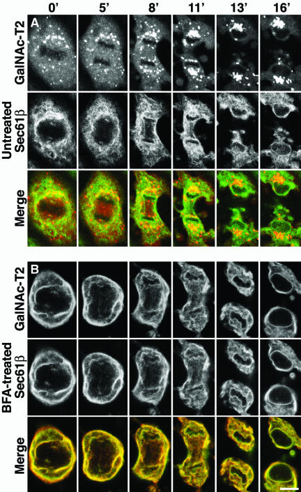
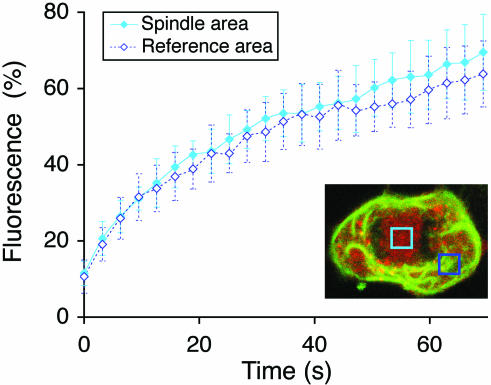
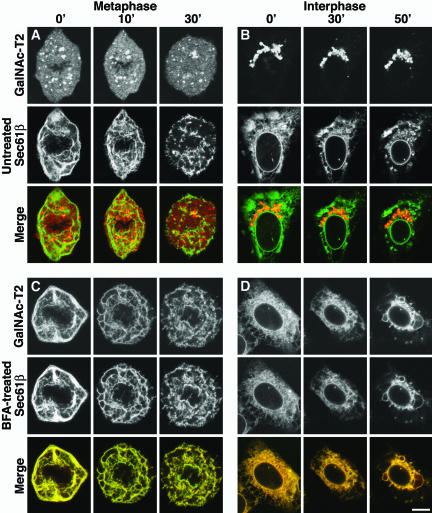
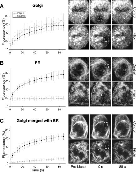
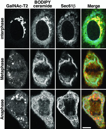
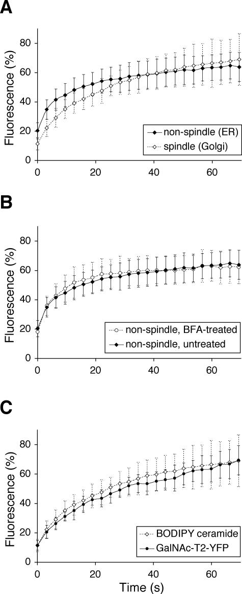
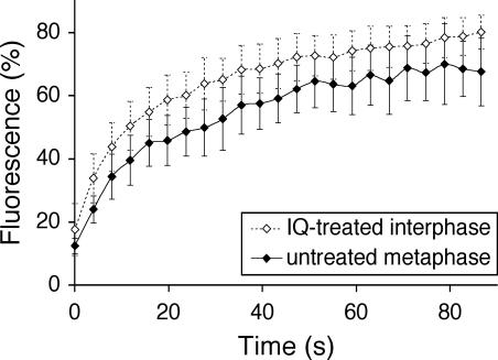
Early in mitosis, the mammalian Golgi apparatus disassembles, and fluorescence microscopy reveals Golgi clusters and an extensive, nonresolvable haze that either represents scattered vesicles or a merged endoplasmic reticulum (ER)-Golgi compartment. To help decide between these alternatives, we have carried out a combined microscopic and pharmacological analysis, by using a BS-C-1 cell line stably coexpressing ER and Golgi markers. Video fluorescence microscopy showed that these two organelles were morphologically distinguishable at all stages of mitosis, and photobleaching experiments showed that diffusion of the Golgi marker was unaffected by the presence of the ER. Fragmentation of the ER by using filipin III completely blocked diffusion of the ER marker but had no effect on the Golgi marker, unless it was first relocated to the ER by using brefeldin A. The Golgi haze was also studied using BODIPY ceramide. Its diffusion was slower in mitotic Golgi than in mitotic ER, but similar to that of a Golgi enzyme marker in the mitotic Golgi haze or in Golgi vesicles generated by ilimaquinone. Together, these results support the idea that the Golgi and the ER remain separate during mitosis and strongly suggest that Golgi markers move by vesicle diffusion, as opposed to lateral diffusion in continuous membranes.


Figures

Figure 1.
Figure 1.
Morphological differences between mitotic ER and Golgi in live cells. BSC-1 cells, stably expressing GalNAc-T2-YFP, a Golgi enzyme marker (red), and CFP-Sec61β, an ER marker (green), were monitored by laser scanning confocal microscopy at 37°C from metaphase to cytokinesis (lapsed time in minutes indicated at the top). (A) Untreated cell. (B) Cell possessing an artificial ER-Golgi hybrid, generated by addition of BFA before M-phase entry (3 h before the first image). Bar, 10 μm.
Figure 2.
Figure 2.
Diffusion rate of the Golgi haze is unaffected by the presence of ER. Anaphase cells were subjected to quantitative FRAP of the Golgi marker GalNAc-T2-YFP (red) both over the ER-free spindle region (filled diamonds) and an ER-containing reference region outside the spindle region (open diamonds). Means ± SD, n = 10. As illustrated in the inset, the absence of ER in the spindle region (light blue box), or presence in the reference region (dark blue box), could be confirmed in every cell imaged by observing the ER marker, CFP-Sec61β.
Figure 3.
Figure 3.
Mitotic and interphase ER can be broken down by using filipin III. Cells were monitored at room temperature (≈20°C) after the addition of filipin III (5 μg/ml) at time points indicated at the top. (A) Metaphase cell. (B) Interphase cell. (C) Metaphase cell, possessing an artificial ER-Golgi hybrid, obtained by addition of BFA before M-phase entry (3 h before the first picture). (D) Interphase cell, likewise treated with BFA for 3 h. Bar, 10 μm.
Figure 4.
Figure 4.
Breakdown of the mitotic ER blocks diffusion of the ER but not the Golgi marker. Metaphase cells were treated with 5 μg/ml filipin III (open diamonds) or left as untreated controls (filled diamonds) for 30-45 min at room temperature, and then subjected to quantitative FRAP (at room temperature). Means ± SD, n = 10. (A) Golgi marker, GalNAc-T2-YFP. (B) ER marker, CFPSec61β. (C) Golgi marker GalNAc-T2-YFP, relocalized to the ER by addition of BFA before M-phase entry (3 h before addition of filipin III). Shown at right are cells representative of each curve (time points at bottom). Boxes indicate bleaching areas (2.5 × 2.5 μm).
Figure 5.
Figure 5.
BODIPY ceramide localizes mostly to the ER in mitotic cells. The GalNAc-T2-YFP (red)/CFP-Sec61β (blue) cell line was loaded with BODIPY ceramide (from a methanol stock; green) for 10 min in the cold, washed, and incubated in fat-free bovine serum albumin for 30 min at 37°C to remove excess dye. Images were sampled, at 37°C, 30-60 min after loading the dye, by using the META system (Carl Zeiss) to separate the spectra. Bar, 10 μm.
Figure 6.
Figure 6.
BODIPY ceramide diffuses more slowly in the mitotic Golgi than in the mitotic ER. Wild-type BS-C-1 cells were stained with BODIPY ceramide as in Figure 5, and 30-60 min after loading the dye, anaphase cells were subjected to quantitative FRAP. (A) Comparison between the ER-free spindle region (open diamonds) and an ER-containing region outside of the spindle (filled diamonds). (B) Comparison between the ER-containing regions of cells that were not (filled diamonds) or were (open circles) pretreated with BFA for 2 h before BODIPY ceramide addition. (C) Comparison between BODIPY ceramide (open diamonds) and GalNAc-T2-YFP (as in Figure 2; filled circles) over the ER-free spindle region. Means ± SD, n = 10.
Figure 7.
Figure 7.
Golgi haze kinetics are consistent with the haze being free vesicles. Quantitative FRAP as for GalNAc-T2-YFP of interphase cells treated for 30-60 min in IQ (open diamonds) and untreated metaphase cells (filled diamonds). Means ± SD, n = 10.

References

    1. Bevis, B.J., Hammond, A.T., Reinke, C.A., and Glick, B.S. (2002). De novo formation of transitional ER sites and Golgi structures in Pichia pastoris. Nat. Cell Biol. 4, 750-756. - PubMed
    1. Birky, C.W. (1983). The partitioning of cytoplasmic organelles at cell division. Int. Rev. Cytol. 15, 49-89. - PubMed
    1. Farmaki, T., Ponnambalam, S., Prescott, A.R., Clausen, H., Tang, B.L., Hong, W., and Lucocq, J.M. (1999). Forward and retrograde trafficking in mitotic animal cells. ER-Golgi transport arrest restricts protein export from the E.R. into COPII-coated structures. J. Cell Sci. 112, 589-600. - PubMed
    1. Glick, B.S., and Malhotra, V. (1998). The curious status of the Golgi apparatus. Cell 95, 883-889. - PubMed
    1. Jesch, S.A., and Linstedt, A.D. (1998). The Golgi and endoplasmic-reticulum remain independent during mitosis in HeLa cells. Mol. Biol. Cell 9, 623-635. - PMC - PubMed

Publication types

MeSH terms

LinkOut - more resources