. 2004 Feb 16;199(4):437-48.
doi: 10.1084/jem.20031457. Epub 2004 Feb 9.

Induction of tumor-specific T cell immunity by anti-DR5 antibody therapy

Affiliations

Induction of tumor-specific T cell immunity by anti-DR5 antibody therapy

Kazuyoshi Takeda et al. J Exp Med. .

Abstract

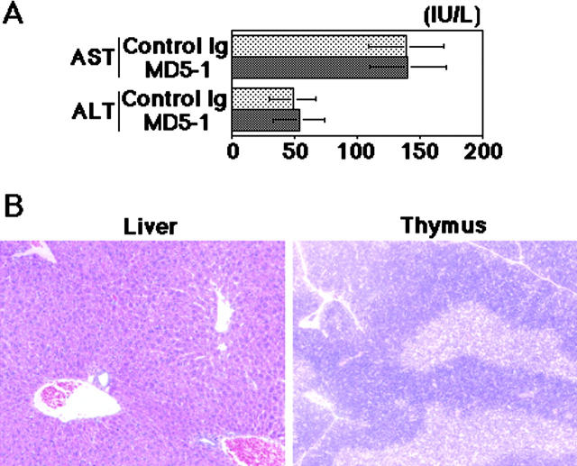
Because tumor necrosis factor-related apoptosis-inducing ligand (TRAIL) preferentially induces apoptosis in tumor cells and plays a critical role in tumor surveillance, its receptor is an attractive target for antibody-mediated tumor therapy. Here we report that a monoclonal antibody (mAb) against the mouse TRAIL receptor, DR5, exhibited potent antitumor effects against TRAIL-sensitive tumor cells in vivo by recruiting Fc receptor-expressing innate immune cells, with no apparent systemic toxicity. Administration of the agonistic anti-DR5 mAb also significantly inhibited experimental and spontaneous tumor metastases. Notably, the anti-DR5 mAb-mediated tumor rejection by innate immune cells efficiently evoked tumor-specific T cell immunity that could also eradicate TRAIL-resistant variants. These results suggested that the antibody-based therapy targeting DR5 is an efficient strategy not only to eliminate TRAIL-sensitive tumor cells, but also to induce tumor-specific T cell memory that affords a long-term protection from tumor recurrence.


Figures

Figure 1.
Figure 1.
Characterization of MD5-1 in vitro. (A) Reactivity with mouse DR5, Fas, and Fn14 transfectants and mouse tumor cell lines. The thick histograms represent MD5-1 staining and the thin histograms represent control Ig staining. (B) Cytotoxic activity of biotinylated MD5-1 against 4T1 cells in the presence (□) or absence (○) of streptavidin. (C) Cytotoxic activity of MD5-1 against 4T1 cells in the presence of P815 cells with (○) or without (□) anti-FcR mAb (2.4G2). (D) Cytotoxic activity of mTRAIL/2PK-3 against 4T1 cells in the presence of MD5-1 or anti-TRAIL mAb (N2B2). (E) Effect of z-VAD-fmk on MD5-1–mediated cytotoxicity against 4T1 tumor cells. (F) Cytotoxic activity of MD5-1 and mTRAIL/2PK3 against R331-mock and R331-FLIP cells. Similar results were obtained in two or three independent experiments.
Figure 2.
Figure 2.
Cytotoxic activity of MD5-1 in the presence of FcR-bearing cells. (A) FcR-mediated cytotoxic activity of MD5-1 against 4T1 tumor cells in the presence of fresh or fixed effector cells. CMA was added to inhibit perforin-dependent cytotoxicity and 2.4G2 was used to block FcγRII and FcγRIII. (B) Cytotoxic activity of MD5-1 against R331-mock and R331-FLIP tumor cells in the presence of various effector cells. (C) Cytotoxic activity of MD5-1 against 4T1 tumor cells in the presence of rabbit or mouse complement.
Figure 3.
Figure 3.
No apparent toxicity of MD5-1 in vivo. (A) Serum AST and ALT levels 20 h after MD5-1 injection. Data are represented as the mean ± SE of 10 mice. Similar results were obtained in two independent experiments at several time points (not depicted). (B) Histological examination (H/E) of liver (×10) and thymus (×5) from MD5-1–treated mice.
Figure 4.
Figure 4.
Contribution of macrophages and NK cells to tumoricidal effect of MD5-1. SCID mice (A) or wild-type BALB/c mice (B) were s.c. inoculated with 4T1 cells, and wild-type BALB/c mice (C) were s.c. inoculated with R331-mock cells. The mice were then treated with MD5-1 (solid symbols) or control Ig (open symbols). Some groups of mice were treated with anti-ASGM1 Ab (triangles), anti-CD11b mAb (circles), or control Igs (squares). (D) Wild-type BALB/c mice were s.c. inoculated with 4T1 cells. The mice were then treated with MD5-1 from day 0 (▪), 15 (•), or 25 (▴). One group of mice was treated with control Ig from day 0 (□) as the control. (E) Wild-type (squares), TRAIL−/− (circles), perforin−/− (triangles), and IFN-γ−/− (upside down triangles) BALB/c mice were s.c. inoculated with R331-mock cells. The mice were then treated with MD5-1 (solid symbols) or control Ig (open symbols). (F) Wild-type (squares), FcRγ−/− (circles), or FcγRII−/− (triangles) BALB/c mice were s.c. inoculated with 4T1 cells. The mice were then treated with MD5-1 (solid symbols) or control Ig (open symbols). Some groups of wild-type mice were treated with both anti-ASGM1 Ab and anti-CD11b mAb (upside down triangles). All data are represented as the mean ± SE of 5–10 mice. Similar results were obtained in two or three independent experiments.
Figure 5.
Figure 5.
MD5-1 induces infiltration of macrophages and DCs in tumor site. Tumor was removed 8 d after tumor inoculation from control Ig-, MD5-1–, or MD5-1– and anti-CD11b mAb (5C6)–treated mice, and then stained with H/E (×5), anti-F4/80 mAb (×10), or anti-CD205 mAb (×10). The marginal region of s.c. tumor is shown. A massive infiltration of F4/80+ macrophages (stained brown) was observed in the marginal region of tumor from MD5-1–treated mice. The arrows are indicated to distinguish CD205+ DCs from nonspecific staining of mast cells. Representative of five tumors in each group.
Figure 6.
Figure 6.
Induction of tumor-specific T cell memory by MD5-1–mediated tumor rejection. (A) BALB/c mice were inoculated with 4T1 cells and then treated with MD5-1 mAb (▪; n = 25) or control Ig (□; n = 5) (left). Simultaneously to secondary challenge with 4T1 cells, some mice were treated with anti-CD4 mAb (•), anti-CD8 mAb (▴), anti-CD4 and anti-CD8 mAbs (▾), or control Ig (▪) (middle). Naive mice were inoculated with 4T1 cells as the control (□). Irrelevant R331-mock cells were inoculated into 4T1-rejecting mice (♦) or naive mice (⋄) (right). (B) BALB/c (squares) and SCID (circles) mice were inoculated with 4T1 cells and then treated with MD5-1 mAb (solid symbols; n = 25) or control Ig (open symbols; n = 5) (left). 5 wk after the first inoculation, the MD5-1–treated BALB/c (▪) and SCID (•) mice that had rejected 4T1 were secondarily inoculated with 4T1 cells (middle). Naive BALB/c (□) and SCID (○) mice were inoculated with 4T1 cells as the control. 2 d after inoculation of 4T1 cells, some SCID mice were i.v. transferred with splenic T cells from the MD5-1–treated BALB/c mice that had rejected 4T1 (•) or naive BALB/c mice (▵) (right). Naive SCID mice (○) and MD5-1–treated wild-type BALB/c mice that had rejected 4T1 (▪) were inoculated with 4T1 cells as the control. (C) Naive BALB/c mice (▿), MD5-1–treated wild-type BALB/c mice that had rejected live 4T1 (□), BALB/c mice immunized by irradiated 4T1 cells precoated with control Ig (▵), or BALB/c mice immunized by irradiated 4T1 cells precoated with MD5-1 (○) were inoculated with the indicated number of live 4T1 tumor cells. (D) Cytotoxic activity of splenic T cells against 4T1 (left) or R331-mock (right) target cells. Effector splenic T cells were prepared from BALB/c mice that had rejected live 4T1 cells by MD5-1 treatment (□), BALB/c mice that had been immunized by irradiated 4T1 cells precoated with MD5-1 (○) or control Ig (▵), or naive BALB/c mice (▿). All data are represented as the mean ± SE of 5–10 mice. Similar results were obtained in three to five independent experiments.
Figure 7.
Figure 7.
Induction of perforin- and FasL-mediated T cell cytotoxicity that can also eliminate TRAIL-resistant variants. (A) BALB/c mice were inoculated with R331-mock (squares) or R331-FLIP (circles) cells, and then treated with MD5-1 mAb (solid symbols; n = 20) or control Ig (open symbols; n = 5) (left). The cured mice were secondarily challenged with R331-mock (▪) or R331-FLIP (•) cells (middle). Naive mice were inoculated with R331-mock (□) or R331-FLIP (○) cells, and the mice preimmunized by irradiated R331-mock cells were inoculated with R331-mock cells (▵) as the controls (middle). Simultaneously to the secondary challenge with R331-mock cells, some mice were treated with anti-CD8 mAb (▵), anti-CD4 and anti-CD8 mAbs (▿), or control Ig (□) (right). (B) R331-FLIP cells were inoculated into the left flank (•) at the indicated time point after R331-mock cell inoculation into the right flank (▪) and commencement of MD5-1 treatment on day 0. As a control, R331-FLIP cells were inoculated into naive mice (○) at the same time as the R331-FLIP inoculation into the treated mice. (C) BALB/c mice that had rejected R331-mock by MD5-1 treatment were secondarily challenged with the indicated number of R331-mock or R331-FLIP cells (○). Naive mice were inoculated with the same numbers of R331-mock or R331-FLIP cells and treated with MD5-1 (▪) or control Ig (□) for comparison. (D) Wild-type (squares) or perforin−/− (circles) BALB/c mice were inoculated with R331-mock cells, and then treated with MD5-1 mAb (solid symbols; n = 20) or control Ig (open symbols; n = 5) (left). The cured mice were secondarily challenged with R331-mock cells. Simultaneously to the secondary challenge, some wild-type (middle) and perforin−/− (right) mice were treated with anti-FasL neutralizing mAb (○), anti-TRAIL neutralizing mAb (▵), anti-FasL and anti-TRAIL mAbs (▿), or control Ig (□). Naive wild-type or perforin−/− mice were inoculated with the same number of R331-mock cells as a control (⋄). All data are represented as the mean ± SE of 5–10 mice. Similar results were obtained in two independent experiments.

References

    1. Wiley, S.R., K. Schooley, P.J. Smolak, W.S. Din, C.P. Huang, J.K. Nicholl, G.R. Sutherland, T.D. Smith, C. Rauch, C.A. Smith, et al. 1995. Identification and characterization of a new member of the TNF family that induces apoptosis. Immunity. 3:673–682. - PubMed
    1. Pitti, R.M., S.A. Marsters, S. Ruppert, C.J. Donahue, A. Moore, and A. Ashkenazi. 1996. Induction of apoptosis by Apo-2 ligand, a new member of the tumor necrosis factor cytokine family. J. Biol. Chem. 271:12687–12690. - PubMed
    1. Nagata, S. 1997. Apoptosis by death factor. Cell. 88:355–365. - PubMed
    1. Takeda, K., Y. Hayakawa, M.J. Smyth, N. Kayagaki, N. Yamaguchi, S. Kakuta, Y. Iwakura, H. Yagita, and K. Okumura. 2001. Involvement of tumor necrosis factor-related apoptosis-inducing ligand in surveillance of tumor metastasis by liver natural killer cells. Nat. Med. 7:94–100. - PubMed
    1. Takeda, K., M.J. Smyth, E. Cretney, Y. Hayakawa, N. Kayagaki, H. Yagita, and K. Okumura. 2002. Critical role for tumor necrosis factor–related apoptosis-inducing ligand in immune surveillance against tumor development. J. Exp. Med. 195:161–169. - PMC - PubMed

Publication types

MeSH terms

Substances