. 2004 Feb 16;199(4):491-502.
doi: 10.1084/jem.20031800. Epub 2004 Feb 9.

B lineage-specific regulation of V(D)J recombinase activity is established in common lymphoid progenitors

Affiliations

B lineage-specific regulation of V(D)J recombinase activity is established in common lymphoid progenitors

Lisa Borghesi et al. J Exp Med. .

Abstract

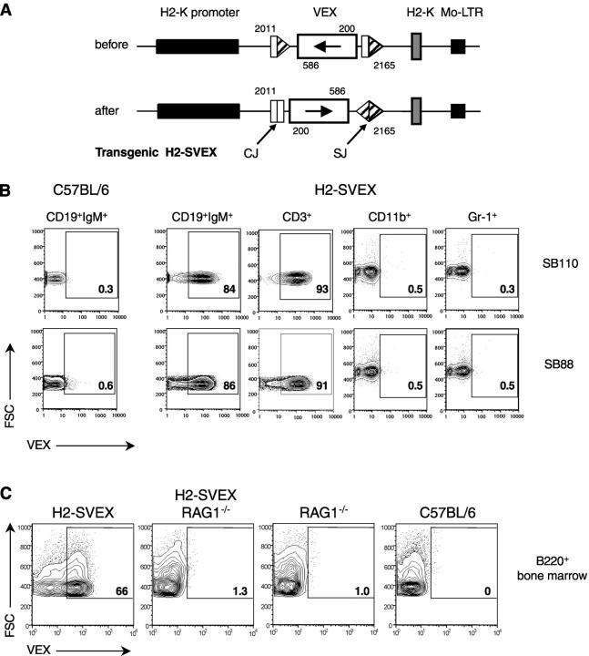
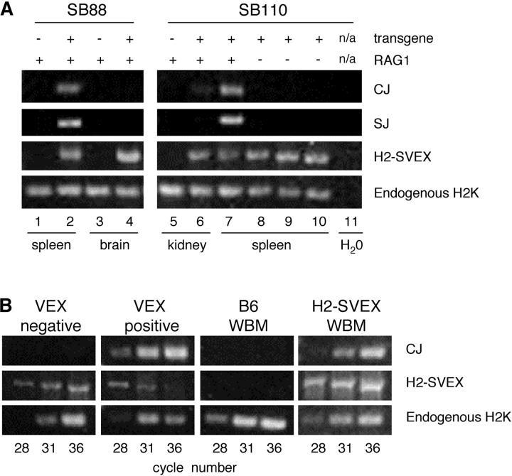
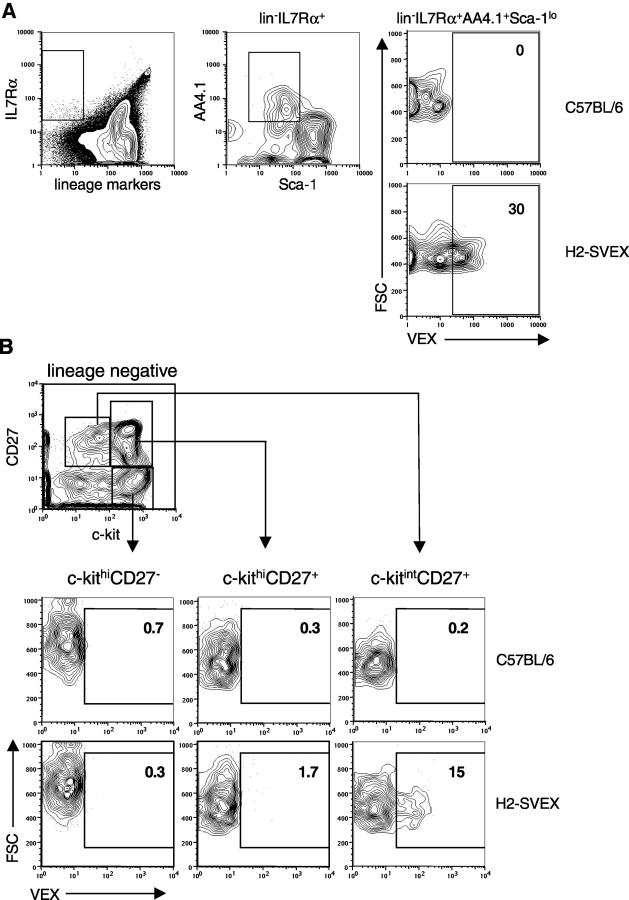
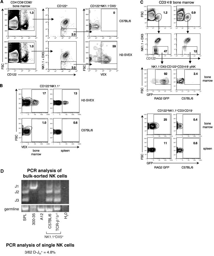
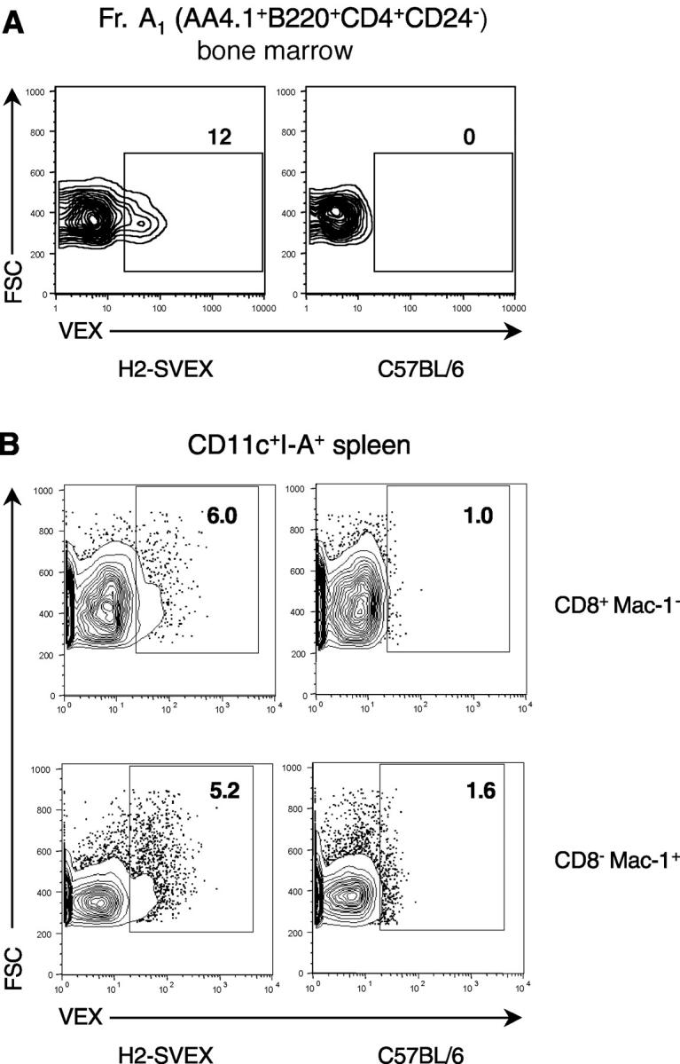
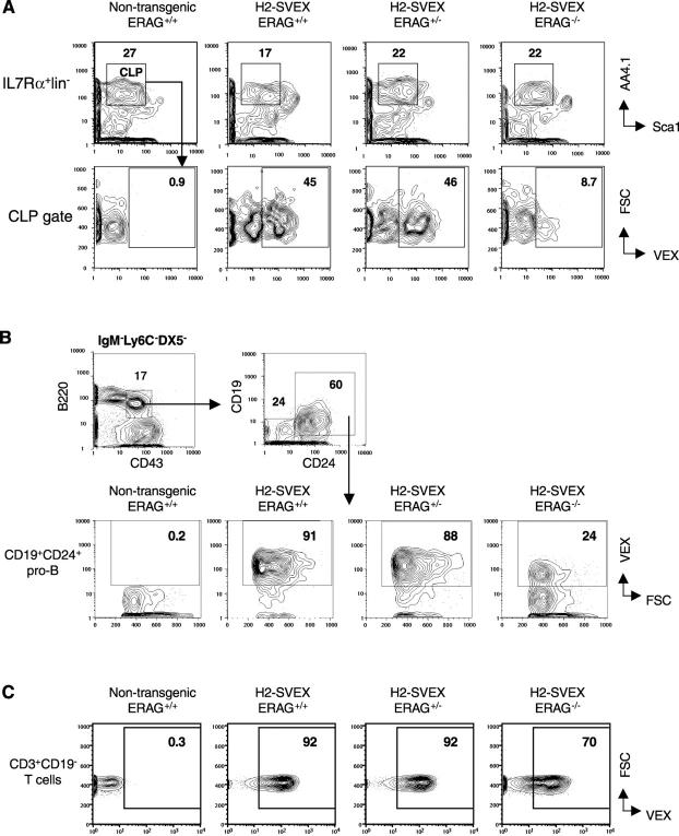
Expression of V(D)J recombinase activity in developing lymphocytes is absolutely required for initiation of V(D)J recombination at antigen receptor loci. However, little is known about when during hematopoietic development the V(D)J recombinase is first active, nor is it known what elements activate the recombinase in multipotent hematopoietic progenitors. Using mice that express a fluorescent transgenic V(D)J recombination reporter, we show that the V(D)J recombinase is active as early as common lymphoid progenitors (CLPs) but not in the upstream progenitors that retain myeloid lineage potential. Evidence of this recombinase activity is detectable in all four progeny lineages (B, T, and NK, and DC), and rag2 levels are the highest in progenitor subsets immediately downstream of the CLP. By single cell PCR, we demonstrate that V(D)J rearrangements are detectable at IgH loci in approximately 5% of splenic natural killer cells. Finally, we show that recombinase activity in CLPs is largely controlled by the Erag enhancer. As activity of the Erag enhancer is restricted to the B cell lineage, this provides the first molecular evidence for establishment of a lineage-specific transcription program in multipotent progenitors.


Figures

Figure 1.
Figure 1.
(A) The transgenic H2-SVEX substrate contains VEX (white rectangle) driven by the murine H2K promoter (black rectangle). VEX within the substrate is initially in the antisense orientation and is flanked by V(D)J recombination signal sequences (triangles) which direct inversional recombination. Primers used to discriminate H2-SVEX before and after rearrangement are indicated (2011, 200, 586, and 2165; as described in Materials and Methods). (B) Splenocytes from SB110 and SB88 H2-SVEX animals were stained with antibodies to detect CD19+ IgM+ B cells, CD3+ T cells, CD11b+CD3CD19NK1.1 myeloid cells, or Gr-1+ CD19CD3NK1.1 granulocytes and subsequently examined for VEX expression. The percentage of VEX+ cells in the gate is given, and outliers are shown. (C) H2-SVEX recombination depends on RAG1. B220+ B cells in the bone marrow were examined for VEX expression in H2-SVEX SB110, H2-SVEX RAG1−/−, RAG−/−, and nontransgenic C57BL/6 control mice. The percentage of VEX+ cells in the gate is given. H2-SVEX RAG1−/− mice were identified by PCR analysis of the SVEX cassette as depicted in A and Fig. 2 A. Identical results were obtained with SB88 H2-SVEX RAG1−/− mice (not depicted). The data presented are representative of six independent experiments.
Figure 2.
Figure 2.
(A) PCR analysis of H2-SVEX V(D)J recombination. H2-SVEX recombination products were examined in two different transgenic lines, SB88 (left panel) or SB110 (right panel), which express or lack RAG as indicated. Nontransgenic C57BL/6 animals are presented as a control. Primers specific to the transgene were used to detect the recombination products CJs and SJs in the indicated tissues. Primers that bind within the murine H2K gene were used to amplify the 1-kb SVEX cassette in transgenic mice (independent of recombination; labeled H2-SVEX in the figure) or to amplify a distinct 250-bp PCR product in both transgenic and wild-type mice (labeled endogenous H2K) derived from the endogenous H2K gene. Amplification of endogenous H2K serves as a positive control confirming the presence of template DNA. (B) Semiquantitative PCR analysis of H2-SVEX recombination. SB88 H2-SVEX bone marrow was FACS® sorted into VEX and VEX+ populations. DNA from an equivalent number of cells was subject to PCR analysis for H2-SVEX transgene-specific CJs, the H2-SVEX transgene, and endogenous H2K. The PCR conditions were designed to give linear amplification for CJs, and cell cycle number is indicated. The data are representative of two to five experiments using cells from independent sorts.
Figure 3.
Figure 3.
V(D)J recombinase activity in multipotent hematopoietic progenitors. (A) V(D)J recombinase activity in CLPs. The lin subset (B220, CD11b, GR-1, Ter119, CD3) of bone marrow obtained from SB88 H2-SVEX transgenic or C57BL/6 control mice was examined for VEX expression in IL-7Rα+AA4.1+Sca-1lo/− cells. (B) The lin subset of bone marrow obtained from SB110 H2-SVEX transgenic or C57BL/6 control mice was examined for VEX expression as a function of c-kit and CD27. The data are representative of two to five independent experiments.
Figure 4.
Figure 4.
Detection of V(D)J recombinase activity in natural killer cells. (A) The CD4CD8CD90 subset of bone marrow was examined for CD122 and DX5 or NK1.1 expression in control B6 (top) or SB110 H2-SVEX transgenic mice (bottom). Immature CD122+DX5 NK1.1 pNK cells (gated population in left and middle) were subsequently analyzed for VEX expression (right). (B) CD122+NK1.1+ cells lacking CD3, CD4, CD8, and CD19 were examined for VEX expression in bone marrow (left) or spleen (right) obtained from SB88 H2-SVEX transgenic (top) or control B6 mice (bottom). Identical results were obtained using SB110 H2-SVEX transgenic mice (not depicted). (C) Bone marrow CD3CD4CD8CD122+NK1.1DX5 pNK (top) and CD3 CD4CD8CD122+NK1.1+CD3CD19 NK (bottom) were examined for GFP expression in RAG2 GFP NG transgenic (left) or control B6 mice (right). The percentage of cells in each gate is given. (D) NK1.1+DX5+CD19CD3CD4CD8 spleen cells were sorted from C57BL/6 or βδ-TCR−/−mice were analyzed by PCR for D-JH joins. PCR analysis of bulk sorted cells is depicted in the gel on the top. SPL refers to total splenic cells. Two cells lines with known rearrangement patterns were used as controls. The AMLV-transformed cell line A12 is derived from a RAG1−/− mouse and therefore retains germline Ig configuration (61). The AMLV-transformed “DJH fixed” 300–35 line bears a D-JH1 rearrangement (62). D-JH4 was not examined. By PCR analysis of single, sorted NK cells from C57BL/6 mice, we determined that the frequency of splenic NK that harbor D-JH rearrangements was 4.8% (3 D-JH + cells out of 62 NK cells analyzed) as indicated on the bottom. The data in A–C are representative of two to five independent experiments. The data in D are representative of at least two experiments using independently sorted cells.
Figure 5.
Figure 5.
Detection of V(D)J recombinase activity in DCs. (A) VEX expression was examined in AA4.1+B220+CD4+CD24 bone marrow progenitors from SB110 H2-SVEX transgenic (left) or B6 control mice (right). (B) Splenic DCs were enriched by collagenase digestion and Nycodenz gradient centrifugation of tissue obtained from SB110 H2-SVEX transgenic (left) or C57BL/6 control mice (right). The CD11c+I-A+ subset was examined for expression of CD8, Mac-1, and VEX. The percentage of cells in each gate is given. The data are representative of at least five (A) or two (B) independent experiments, and identical results were obtained with SB88 H2-SVEX transgenics (not depicted).
Figure 6.
Figure 6.
Regulation of V(D)J recombinase activity in multipotential lineage progenitors. (A) Bone marrow from SB110 H2-SVEX animals that were Erag +/+, Erag +/−, or Erag −/− was stained for IL-7Rα+AA4.1+Sca-1lo/−lin CLPs (top), and cells within the CLP gate were examined for VEX expression (bottom). Identical results were obtained when wild-type SB110 and SB88 H2-SVEX mice were compared (not depicted). (B) VEX expression was analyzed in B220+CD43+DX5Ly6CIgM CD19+CD24+ pro-B cells (bottom row). (C) VEX expression was analyzed in CD3+CD19 splenic T cells. The percentage of each population within the gate is given. The data are representative of two to five independent experiments.

Similar articles

Cited by

References

    1. Nutt, S.L., D. Eberhard, M. Horcher, A.G. Rolink, and M. Busslinger. 2001. Pax5 determines the identity of B cells from the beginning to the end of B-lymphopoiesis. Int. Rev. Immunol. 20:65–82. - PubMed
    1. Rothenberg, E.V., and C.J. Dionne. 2002. Lineage plasticity and commitment in T-cell development. Immunol. Rev. 187:96–115. - PubMed
    1. Mombaerts, P., J. Iacomini, R.S. Johnson, K. Herrup, S. Tonegawa, and V.E. Papaioannou. 1992. RAG-1-deficient mice have no mature B and T lymphocytes. Cell. 68:869–877. - PubMed
    1. Shinkai, Y., G. Rathbun, K.P. Lam, E.M. Oltz, V. Stewart, M. Mendelsohn, J. Charron, M. Datta, F. Young, A.M. Stall, et al. 1992. RAG-2-deficient mice lack mature lymphocytes owing to inability to initiate V(D)J rearrangement. Cell. 68:855–867. - PubMed
    1. Gao, Y., J. Chaudhuri, C. Zhu, L. Davidson, D.T. Weaver, and F.W. Alt. 1998. A targeted DNA-PKcs-null mutation reveals DNA-PK-independent functions for KU in V(D)J recombination. Immunity. 9:367–376. - PubMed

Publication types