. 2004 Feb 16;164(4):581-91.
doi: 10.1083/jcb.200307176. Epub 2004 Feb 9.

In Candida albicans, the Nim1 kinases Gin4 and Hsl1 negatively regulate pseudohypha formation and Gin4 also controls septin organization

Affiliations

In Candida albicans, the Nim1 kinases Gin4 and Hsl1 negatively regulate pseudohypha formation and Gin4 also controls septin organization

Raymond Wightman et al. J Cell Biol. .

Abstract

In the development of hyphal germ tubes of Candida albicans, a band of septin forms at the base of the germ tube (basal septin band). Later, a septin ring forms, which organizes the first septum within the germ tube (septin ring). We have investigated the role of the Nim1 kinases, Gin4 and Hsl1, in the formation of these septin structures. We show that during germ tube formation, Gin4 is required for the organization of the septin ring but not the basal septin band. Hsl1 is not required for the formation of either septin rings or basal bands. Unexpectedly, we found that both gin4Delta and hsl1Delta mutants form pseudohyphae constitutively, in a fashion that in the case of gin4Delta, is partly independent of Swe1. Gin4-depleted pseudohyphae are unable to form hyphae when challenged with serum, but this can be overcome by ectopic expression of Gin4 from the MET3 promoter. Thus, Gin4 may regulate the developmental switch from pseudohyphae to hyphae.


Figures

Figure 1.
Figure 1.
Structural comparisons of the C. albican s Nim1 kinase homologues with S. cerevisiae Gin4 and Hsl1. Unique motifs shared between homologues are shown for Gin4 (solid gray) and Hsl1 (solid black). For detailed alignments see Fig. S1. PK, protein kinase domain (black diagonals); Motif 1 (TYA); Motif 2 (SQL); Motif 3 (GSFFRK); Motif 4 (VVQSVXXATKRLS).
Figure 2.
Figure 2.
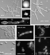
The phenotype of gin4Δ and MET3-GIN4 cells. MET3-GIN4 and gin4Δ strains were grown on YEPD at 30°C and the appearance of colonies and cells were recorded as indicated. Chitin was visualized with calcofluor white and septin by immunocytofluorescence using αCdc11 antiserum (Sudbery, 2001). “MET3-GIN4 off/on” refers to whether the MET3 promoter was repressed or derepressed. When repressed, cells were first grown in synthetic-defined medium under derepressing conditions and then grown for 6 h in derepressing conditions. Note: septin and chitin rings, indicated by arrows with solid heads in g and h, formed during the growth in derepressing conditions. The round patches of septin within the cells (h, open arrows) colocalized with nuclei as revealed by DAPI staining (not depicted). Panels a and d are shown at the same magnification. Bars, 10 μm. Panels e–k are also shown at the same magnification.
Figure 3.
Figure 3.
gin4Δ cells are hyperinvasive. (a) Wild-type (BWP17) and gin4Δ cells were grown on YEPD agar plates for 7 d. Colonies were photographed before and after the plates had been washed. (b) Wild-type and gin4Δ cells were patched over an area of a YEPD plate or a YEPD plate containing 20% serum and incubated at 30°C or 37°C, as indicated, and incubated for 7 d. After washing, transverse sections were cut from the plate and examined using a dissecting microscope.
Figure 4.
Figure 4.
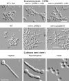
The C. albicans SWE 1 gene. (a–f) S. cerevisiae cells of the indicated genotype were grown for 16 h in the presence of galactose to induce the GAL1 promoter. IAA, isoamyl alcohol. (g–i) C. albicans swe1Δ cells were grown to stationary phase as yeast and reinoculated for 2 h to form hyphae (g), pseudohyphae (h), or yeast (i). Bars, 10 μm.
Figure 5.
Figure 5.
A swe1Δ Gin4-depleted cell mutant still forms filaments. (a and b) Cells of the indicated genotype were grown to stationary phase in derepressing conditions and reinoculated into fresh medium in repressing conditions. After 6 h, cells were stained with DAPI (blue) and images were recorded using DIC optics. (c) MET3-GIN4 and swe1Δ MET3-GIN4 strains were grown for 3 d at 30°C under MET3-repressing conditions as indicated. (d) The same plates were incubated for a further 2 d and examined for evidence of agar invasion by the plate washing assay described in Fig. 3.
Figure 6.
Figure 6.
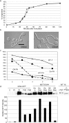
Gin4-depleted cells can form hyphae but only from the yeast state. (a and b) MET3–GIN4 cells were grown to stationary phase under derepressing conditions. The resulting unbudded cells were reinoculated into derepressing (GIN4 on) or repressing (GIN4 off) medium containing 20% serum. (a) At the indicated times the percentage evagination was recorded. Solid squares, GIN4 on; solid triangles, GIN4 off. Each datum is the mean of at least 80 cells. (b) The appearance of hyphae recorded after 120 min using DIC optics. Bar, 10 μm. (c) MET3-GIN4 cells were grown to stationary phase in derepressing medium. Cells were then reinoculated, at a concentration of 105 cells/ml−1, into MET3 derepressing (“On-PH”) or repressing (“Off-PH”) medium, pH 6, 36°C, which promotes the formation of pseudohyphae. At 1-h intervals, samples were withdrawn, and cells were collected by centrifugation and resuspended into the same volume of fresh, prewarmed, derepressing or repressing medium containing 20% serum. After a further 2-h incubation, the proportion of elongated cells producing hyphae was recorded. Elongated cells are defined as cells in which the length was more than twice the width. It should be noted that at early time points, some round cells persisted and that a high proportion of these produced germ tubes, which were not included in the analysis shown in Fig. 6 c. However, by the 6-h time point, <5% of the population were round cells. Parallel samples of the parental BWP17 strain were grown to stationary phase in repressing conditions and incubated either at 30°C, pH 6, which induced yeast growth (WT-Y) or pseudohyphal inducing conditions (WT-PH) before serum treatment to induce hyphae. (d) The experiment described in c was repeated with GIN4-GFP (GIN4 expressed from its own promoter) grown under MET3-repressing conditions and MET3-GIN4-GFP strains grown under repressing (“Off”) or derepressing conditions (“On”). Cells were grown as yeast (Y) or pseudohyphae (PH) and samples were withdrawn 2 and 6 h as indicated (Y2, Y6, PH2, PH6). Gin4 levels were determined by Western blotting. Quantification is by direct measurement of ECL. Full details, including normalization of loading, are given in supplemental material. The protein sample from wild-type cells grown as yeast for 6 h (Y6) was treated with 1,000 U λ phosphatase (New England Biolabs, Inc.) or an equivalent volume of water. The loading was reduced to show the band shift clearly. After withdrawal of the samples for Western blotting, the remaining cultures were treated with serum to induce germ tubes. Quantitation of the proportion of each culture producing germ tubes is shown in Table S2.
Figure 7.
Figure 7.
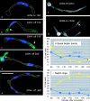
Properties of gin4Δ hyphae. MET3GIN4 cells were grown to stationary phase under derepressing conditions. The resulting unbudded cells were reinoculated into derepressing (GIN4 on) or repressing (GIN4 off) medium containing 20% serum and stained with αCdc11 antibody (a–e, false color green) and DAPI (a–e, false color blue) or calcofluor (f and g). Arrows in b and d indicate examples of a basal septin band. Arrow in c indicates apical staining. Arrow in g indicates primary septum. All panels are at the same magnification. Bar, 10 μm. (h and i) Quantitation of cells with tubes and septin bands. A minimum of 70 cells was counted for each time point.
Figure 8.
Figure 8.
Properties of hyphae dependent on the ME T3 regulated expression of GIN4-GFP. MET3-GIN4 cells were grown to stationary phase in derepressing medium, and then reinoculated into fresh medium containing serum at 37°C in repressing or derepressing conditions as described in Fig. 7. The percentage of evagination, germ tubes with septin rings, basal septin bands, and two nuclei were recorded. A minimum of 50 cells were counted for each time point. Levels of Gin4-GFP were monitored during the time course by Western blot analysis (e) and quantified (f) as described in supplemental material. Arrows indicate the Gin4-GFP band. The faint band beneath the Gin4 band is a nonspecific band present in non-GFP tagged strains. We have also used this as a loading control, with similar results to those presented.
Figure 9.
Figure 9.
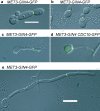
Gin4-GFP does not form a ring at the base of germ tubes. MET3-GIN4-GFP or MET3-GIN4 CDC10-GFP, as indicated, were grown in YEPD medium, pH 6.0, as follows: (a) 37°C; (b) 30°C; (c and d) 37°C plus serum (70 min); and (e) 37°C plus serum (210 min). Bars, 10 μm.
Figure 10.
Figure 10.
hsl1Δ mutants form pseudohyphae constitutively despite forming normal septin rings and septa. The C. albicans hsl1Δ strain was grown in YEPD at 30°C to an OD600 = 0.6. Panel and images recorded as indicated. (b) Immunocytofluorescence was performed using S. cerevisiae αCdc11 antisera and DAPI staining to reveal nuclei. Arrow indicates a septin ring; N = nuclei. Panels c and d were captured with a DeltaVision microscope, the fields correspond to the area within dotted lines in a. (d) A three-dimensional reconstruction of the image rotated 140° around the x axis with respect to the image in c. Arrows with solid heads in c and d indicate a ring of chitin at the base of the growing cells. The open arrow shows a fully formed primary septum. (e) hsl1Δ was grown to high cell density (OD600 = 0.75) to promote the formation of yeast cells. A sample was diluted into fresh YEPD plus serum medium and incubated for 2 h at 37°C. Cells were fixed and stained with anti-Cdc11 antibody as described previously (Sudbery, 2001). Bars, 5 μm.

References

    1. Ahn, S.H., A. Acurio, and S.J. Kron. 1999. Regulation of G2/M progression by the STE mitogen-activated protein kinase pathway in budding yeast filamentous growth. Mol. Biol. Cell. 10:3301–3316. - PMC - PubMed
    1. Altman, R., and D. Kellogg. 1997. Control of mitotic events by Nap1 and the Gin4 kinase. J. Cell Biol. 138:119–130. - PMC - PubMed
    1. Barral, Y., M. Parra, S. Bidlingmaier, and M. Snyder. 1999. Nim1-related kinases coordinate cell cycle progression with the organization of the peripheral cytoskeleton in yeast. Genes Dev. 13:176–187. - PMC - PubMed
    1. Berman, J., and P.E. Sudbery. 2002. Candida albicans: A molecular revolution built on lessons from budding yeast. Nat. Rev. Genet. 3:918–930. - PubMed
    1. Booher, R.N., R.J. Deschenes, and M. Kirschner. 1993. Properties of Saccharomyces cerevisiae wee1 and its differential regulation in response to G1 and G2 cyclins. EMBO J. 12:3417–3428. - PMC - PubMed

Publication types

MeSH terms