. 2004 Feb 17;101(7):1922-6.
doi: 10.1073/pnas.0308693100. Epub 2004 Feb 9.

Ceramidase expression facilitates membrane turnover and endocytosis of rhodopsin in photoreceptors

Affiliations

Ceramidase expression facilitates membrane turnover and endocytosis of rhodopsin in photoreceptors

Usha Acharya et al. Proc Natl Acad Sci U S A. .

Abstract

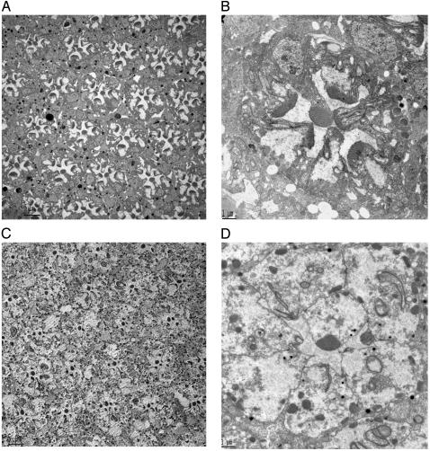
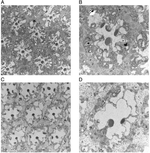
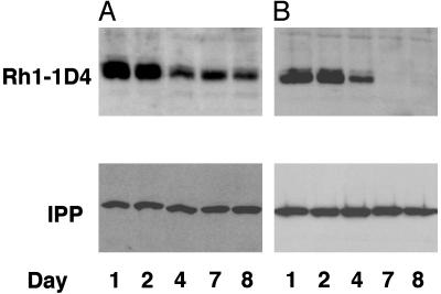
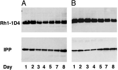
Transgenic expression of ceramidase suppresses retinal degeneration in Drosophila arrestin and phospholipase C mutants. Here, we show that expression of ceramidase facilitates the dissolution of incompletely formed and inappropriately located elements of rhabdomeric membranes in ninaE(I17) mutants lacking the G protein receptor Rh1 in R1-R6 photoreceptor cells. Ceramidase expression facilitates the endocytic turnover of Rh1. Although ceramidase expression aids the removal of internalized rhodopsin, it does not affect the turnover of Rh1 in photoreceptors maintained in dark, where Rh1 is not activated and thus has a slower turnover and a long half-life. Therefore, the phenotypic consequence of ceramidase expression in photoreceptors is caused by facilitation of endocytosis. This study provides mechanistic insight into the sphingolipid biosynthetic pathway-mediated modulation of endocytosis and suppression of retinal degeneration.


Figures

Fig. 1.
Fig. 1.
Expression of ceramidase clears rhabdomeric remnants in ninaEI17 mutant photoreceptors. (A) ninaEI17 mutants lacking Rh1 in R1–R6 photoreceptors have defects in rhabdomere biogenesis. A cross section of a compound eye is shown. In transmission electron microscopy sections of 3-day-old fly eyes, all ommatidial units display degenerative rhabdomeres in R1–R6 photoreceptors, and rhabdomeric membranes are visible in the photoreceptor cell body. (Scale bar, 5 μm.) (B) A higher-magnification view across a single ommatidium of 3-day-old ninaEI17 shows that rhabdomeres are incompletely formed, and rhabdomeric elements are seen involuting into the photoreceptors in R1–R6 cells. (Scale bar, 1 μm.) (C) A low-magnification view of 3-day-old ninaEI17 flies expressing ceramidase. In this section, R1–R6 photoreceptors in most ommatidial units have reduced or no rhabdomere, and discrete tubulovesicular structures appear intracellularly. (Scale bar, 5 μm.) (D) A high-magnification view of one of the ommatidial units from C shows that most of the involuting rhabdomeric membranes are missing in R1–R6 cells. Some membranous structures in the cell are seen connected to the apical plasma membrane. Distinct tubulovesicular structures are also seen in the cell body. (Scale bar, 1 μm.)
Fig. 2.
Fig. 2.
Ceramidase facilitates the removal of rhabdomeric elements when expressed in adult photoreceptors. (A) Transmission electron microscopy of ninaEI17 ommatidium subjected to heat shock for 3 days at 37°C for 1 h/day. The heat shock was induced from day 1 to day 3 after eclosion, and the compound eyes were examined on day 4. Heat shock resulted in slight compacting of incompletely formed rhabdomeres. However, involuting rhabdomeric elements were still distinguished in these mutant photoreceptors. (Scale bar, 2 μm.) (B) Higher-magnification view of one ommatidium from ninaEI17 photoreceptors treated as described in A. (Scale bar, 1 μm.) (C) TEM of ninaEI17 with ceramidase photoreceptors subjected to heat shock for 3 days at 37°C for 1 h/day to induce ceramidase expression. Heat shock was induced from day 1 to day 3, and eyes were examined on day 4. Induction of ceramidase resulted in dissolution of rhabdomeric elements and clearing of the photoreceptors. (Scale bar, 2 μm.) (D) Higher-magnification of a single ommatidial unit from photoreceptors shown in C. (Scale bar, 1 μm.)
Fig. 3.
Fig. 3.
Ceramidase expression facilitates the endocytic turnover of Rh1. (A) A pulse of 1D4-tagged Rh1 was induced in w1118 control flies by heat shock for 1 h at 37°C, and proteins were extracted from these fly retinas on days 1, 2, 4, 7, and 8 and were analyzed by Western blot for the tagged Rh1. The Rh1 level peaked between 1 and 2 days and decreased in amount thereafter, and a signal was still detected on day 8. (B) A pulse of 1D4-tagged Rh1 was induced as in A in flies expressing UAS-ceramidase under a GMR-GAL4 driver. In these flies the Rh1 peaked as in control retina, however, the tagged Rh1 was not visible beyond day 4. The blots were probed for IPP as loading controls (Lower).
Fig. 4.
Fig. 4.
Ceramidase expression does not facilitate Rh1 turnover in flies maintained in the dark. (A) A pulse of 1D4-tagged Rh1 was induced in w1118 control flies by heat shock for 1 h at 37°C, and proteins were extracted from retina 1, 2, 3, 4, 5, 7, and 8 days after heat shock and were analyzed by Western blot for the tagged Rh1. The flies were maintained in dark during the whole procedure until homogenization. Rh1 turned over slowly in flies maintained in the dark and had a longer half-life, and a substantial fraction was seen even on day 8. (B) A pulse of 1D4-tagged Rh1 was induced in flies expressing UAS-ceramidase under GMR-GAL4 and was analyzed as described in A. The rate of turnover was not enhanced as in the case of light, and a significant fraction of Rh1 was seen even on day 8. The blots were probed for IPP as loading controls (Lower).

References

    1. Merrill, A. H., Jr., Schmelz, E. M., Dillehay, D. L., Spiegel, S., Shayman, J. A., Schroeder, J. J., Riley, R. T., Voss, K. A. & Wang, E. (1997) Toxicol. Appl. Pharmacol. 142, 208–225. - PubMed
    1. van Blitterswijk, W. J., van der Luit, A. H., Veldman, R. J., Verheij, M. & Borst, J. (2003) Biochem. J. 369, 199–211. - PMC - PubMed
    1. Hannun, Y. A., Luberto, C. & Argraves, K. M. (2001) Biochemistry 40, 4893–4903. - PubMed
    1. Acharya, U., Patel, S., Koundakjian, E., Nagashima, K., Han, X. & Acharya, J. K. (2003) Science 299, 1740–1743. - PubMed
    1. Zuker, C. S., Cowman, A. F. & Rubin, G. M. (1985) Cell 40, 851–858. - PubMed

MeSH terms

LinkOut - more resources