. 2004 Feb 17;101(7):1951-6.
doi: 10.1073/pnas.0307145101. Epub 2004 Feb 9.

Mimicking live flavivirus immunization with a noninfectious RNA vaccine

Affiliations

Mimicking live flavivirus immunization with a noninfectious RNA vaccine

Regina M Kofler et al. Proc Natl Acad Sci U S A. .

Abstract

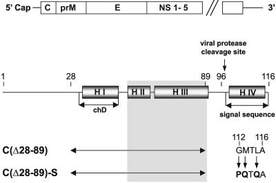
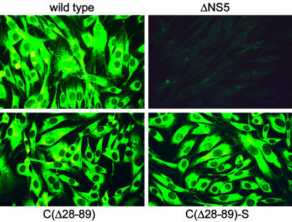
Flaviviruses are human pathogens of world-wide medical importance. They have recently received much additional attention because of their spread to new regions (such as West Nile virus to North America), highlighting their potential as newly emerging disease agents. Using tick-borne encephalitis virus, we have developed and evaluated in mice a new genetic vaccine based on self-replicating but noninfectious RNA. This RNA contains all of the necessary genetic information for establishing its replication machinery in the host cell, thus mimicking a natural infection. However, genetic modifications in the region encoding the capsid protein simultaneously prevent the assembly of infectious virus particles and promote the secretion of noninfectious subviral particles that elicit neutralizing antibodies. These characteristics demonstrate that a new generation of flavivirus vaccines can be designed that stimulate the same spectrum of innate and specific immune responses as a live vaccine but have the safety features of an inactivated vaccine.


Figures

Fig. 1.
Fig. 1.
Schematic of the TBEV genome and protein C. The genome (Upper) consists of a single long ORF encoding three structural proteins (C, prM, and E) and several nonstructural proteins and two flanking noncoding sequences (not drawn to scale). Protein C (Lower) is largely α-helical (four predicted helices, HI to H IV). H I coincides approximately with a stretch of hydrophobic amino acid residues (referred to as central hydrophobic domain, chD). The engineered deletion (double-headed arrow) removes all of H I/chD and an adjacent domain in which compensating mutations restoring viability of deletion mutants were previously observed to arise (shown as shaded area). H IV is an internal signal sequence of the subsequent component of the polyprotein, protein prM. It is cleaved off from mature protein C by the action of the viral protease NS2B/3. In mutant C(▵28–89)-S the signal sequence was modified by three point mutations as indicated in the figure, creating an idealized sequence. Numbers refer to amino acid positions in protein C.
Fig. 2.
Fig. 2.
Expression of viral protein in BHK-21 cells. In vitro transcribed wild-type or mutant RNA (as indicated) was introduced by electroporation, and viral protein expression was determined by immunofluorescence staining with a polyclonal anti-TBEV serum 48 h after transfection. In the absence of RNA replication (replication-deficient mutant ▵NS5) no protein expression was detected.
Fig. 3.
Fig. 3.
Analysis of secreted particles. (A) The kinetics of the release of protein E from BHK-21 cells transfected with wild-type or mutant RNAs was monitored by ELISA. (B) Particles secreted by mutant C(▵28–89)-S were analyzed on a discontinuous (10%, 35%, and 50% as indicated below) sucrose gradient and compared with virus and RSP controls. (C) The buoyant density of particles secreted by mutant C(▵28–89)-S was determined by equilibrium sucrose gradient centrifugation in comparison with RSPs. (D) SDS/PAGE of viral and subviral particles. Particles secreted from cells transfected with C(▵28–89)-S were fractionated and compared with RSP and wild-type virus preparations. The positions of the structural proteins E, M (and its precursor, prM), and C are indicated.

References

    1. Lindenbach, B. D. & Rice, C. M. (2001) in Fields Virology, eds. Knipe, D. M., Howley, P. M., et al. (Lippincott Williams & Wilkins, Philadelphia), pp. 991–1041.
    1. Pugachev, K. V., Guirakhoo, F., Trent, D. W. & Monath, T. P. (2003) Int. J. Parasitol. 33, 567–582. - PubMed
    1. Murphy, B. R. & Chanock, R. M. (2001) in Fields Virology, eds. Knipe, D. M., Howley, P. M., et al. (Lippincott Williams & Wilkins, Philadelphia), pp. 435–467.
    1. Leitner, W. W., Hwang, L. N., deVeer, M. J., Zhou, A., Silverman, R. H., Williams, B. R., Dubensky, T. W., Ying, H. & Restifo, N. P. (2003) Nat. Med. 9, 33–39. - PMC - PubMed
    1. Monath, T. P., Guirakhoo, F., Nichols, R., Yoksan, S., Schrader, R., Murphy, C., Blum, P., Woodward, S., McCarthy, K., Mathis, D. et al. (2003) J. Infect. Dis. 188, 1213–1230. - PubMed

MeSH terms