. 1951 Mar;14(2):133-46.
doi: 10.1152/jn.1951.14.2.133.

Organization of the diffuse thalamic projection system

Organization of the diffuse thalamic projection system

T E STARZL et al. J Neurophysiol. 1951 Mar.
No abstract available


Figures

FIG. 1
FIG. 1
Recruiting responses (records A–H, at left) and spike-wave complexes (records I–Q, at right) evoked by low frequency stimulation of thalamic sites indicated by appropriate symbols on transverse sections through diencephalon (levels A–E, center). Channels record activity from anterior (A) middle (M) and posterior (P) regions of cortex, homolateral (H) or contralateral (C) to side of stimulation, and occasionally from caudate nucleus (NC). Responses were elicited from Stimulating anterior reticular nucleus (records A, B), antero-medial (C), ventralis anterior (D, E, I, J, L), centralis medialis (records F, G), centre median (H), the ventralis medialis (M), ventralis lateralis (O), medial geniculate (P), lateralis posterior (Q) and internal capsule (K, N). Anesthesias employed were: nembutal(records A, L, G, I, M, N, P, Q), chlorolosane (B, D, J, K), beta-erythroidine (F, O) and encephalé isolé (E, H, L). Stimulus voltages varied between 2 and 7, and were usually 5. Legends on diencephalon are: A—anterior nuclei, BP—basis pedunculi, C—centralis medialis, CA—caudate, CL—centralis lateralis, CLA—Claustrum, CM—Centre median, F—fornix, GP—globus pallidus, H—habenula, HP—habenulo-peduncular tract, HV—hypothalamic ventromedial nucleus, IV—internal capsule, L—lateralis posterior, LA—lateralis anterior, LG—lateral geniculate, M—medial nucleus, MB—mammillary body, MG—medial geniculate, OT—optic tract, P—pulvinar, PU—putamen, R—reticular nucleus, SU—subthalamus, VA—ventralis anterior, VL—ventralis lateralis, VM—ventralis medialis, VP—ventralis posterior. Dots indicate diencephalic sites whose stimulations did not evoke wave responses in cortex. Vertical calibration—l00 µV; horizontal calibration—1 sec.
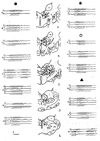
FIG. 2
FIG. 2
Records of turnover of recruiting potentials on passing through claustrum and orbital gyrus (A), cingulate gyrus (B), caudate nucleus (C) and ventralis anterior (D). In each instance, pickups were from responding region and from anterior cortex (HA) on side of stimulation. Stimuli were applied to intralaminar nucleus (A) and ventralis anterior (B–D), under nembutal anesthesia (A)or in encephalé iso1é (B–D).
FIG. 3
FIG. 3
A, B—Records showing spontaneous, low voltage, fast activity recorded under nembutal anesthesia from cortical areas yielding recruiting responses. A: gyrus cinguli showing initial fast activity and its later disappearance. B: orbital gyrus, with fast activity at site displaying largest recruiting response (top channel). Records from anterior cortex (HA) are included for comparison. Stimuli to intralaminar nucleus at 1.5 (A, left) and 7 volts (A, right, and B). Note spontaneous burst at B at site with best fast activity (top channel). C: recruiting responses in anterior hemisphere (HA) and cingulate gyrus (HGC) abolished (lower record) by cut between these structures and thalamus. D: recruiting responses in two regions of orbital gyrus (HOG) abolished (lower record) by cut between this gyrus and thalamus.
FIG. 4
FIG. 4
Records from indicated regions and anterior cortex (HA) showing recruiting responses (left), spontaneous spindle bursts under barbiturate anesthesia (middle) and rhythmic after-discharge following single shock stimuli (right) to intralaminar nuclei (A, E), ventralis anterior (B, C, D), and anterior reticular nucleus (F, G). Stimulating voltages between 1.5 and 7.
FIG. 5
FIG. 5
Transverse sections through hemisphere with shading indicating sites from which recruiting waves may be recorded with deep pickups upon low frequency thalamic stimulation. More darkly shaded regions are those exhibiting rhythmic after-discharge upon single shock stimuli to thalamic recruiting nuclei. Structures displaying spontaneous spindle bursts under nembutal anesthesia have essentially same distribution. Abbreviations are as follows; A—aquedust, AC—anterior commissure, AD—anterodorsal nucleus, AM—-anteromedial nucleus, AMY—amygdala, BP—basis pedunculi, C—nucleus centralis medialis, CA—caudate, CC—corpus callosum, CL—nucleus centralis lateralis, CLA—claustrum, CM—centre median, F—fornix, GAEC—anterior ectosylvian gyrus, GAS—anterior sigmoid gyrus, GASS—anterior suprasylvian gyrus, GASY—anterior sylvian gyrus, GC—cingulate gyrus, GCO—coronal gyrus, GO—lateral gyrus, GMEC—middle ectosylvian gyrus, GMSS—middle suprasylvian gyrus, GO—orbital gyrus, GP—globus pallidus, GPS—posterior sigmoid gyrus, GSP—suprasplenial gyrus, H—hippocampus, HA-habenula, HP—habenulo-peduncular tract, IC—internal capsule, LA—nucleus lateralis anterior, LG—lateral geniculate body, LP—lateralis posterior, M—medial nucleus, MB—mammillary body, MG—medial geniculate body, OB—olfactory bulb, OT—optic tract, P—pulvinar, PC—posterior commissure, PL—pyriform lobe, PRE—pretectal region, PU—putamen, R—reticular nucleus, RN—red nucleus, RS—rhinal sulcus, S—septum, SN—substantia nigra, SU—subthalamus, TEG—midbrain tegmentum, VA—nucleus ventralis anterior, VL—nucleus ventralis lateralis, VM—nucleus ventralis medialis, VP—nucleus ventralis posterior.
FIG. 6
FIG. 6
Views of lateral (A) and medial (B) aspects of hemisphere of cat, with cortical regions from which recruiting responses may be recorded shown by cross-lining. More darkly shaded zones indicate most responsive areas. In C, position of sensory-receiving areas (stipple) and motor cortex (black) has been indicated on lateral aspect of hemisphere, after Rose, and Woolsey (12). Abbreviations are: ASG—anterior sigmoid gyrus, AUD—auditory receiving area, CC—corpus callosum, CING—cingulate gyrus, COR—coronal gyrus, LAT—lateral gyrus, MOT—motor area, MSS—middle suprasylvian gyrus, ORB—orbital gyrus, PRO—gyrus proreus, PSS—posterior suprasylvian gyrus, SOM—somatic receiving area, VIS—visual receiving area.
FIG. 7
FIG. 7
Transverse sections through hemisphere of cat, upon which are indicated origins (black) and projections (stipple) of diffuse thalamic projection system. At right, records illustrate recruiting response to low frequency thalamic stimulation, spindle bursts in barbiturate anesthesia, and rhythmic after-discharge following single shock stimuli to thalamus, exhibited by all regions of projection.

References

    1. Dempsey EW, Morison RS. The production of rhythmically recurrent cortical potentials after localized thalamic stimulation. Amer. J. Physiol. 1942;135:293–300.
    1. Dempsey EW, Morison RS. The interaction of certain spontaneous and induced cortical potentials. Amer. J. Physiol. 1942;135:301–308.
    1. Dempsey EW, Morison RS. The electrical activity of a thalamocortical relay system. Amer. J. Physiol. 1943;138:283–296.
    1. Hunter J, Jasper HH. Effects of thalamic stimulation in unanesthetized animals. EEG clin. Neurophysiol. 1949;1:305–324. - PubMed
    1. Jasper HH, Droogleever-Fortuyn J. Experimental studies on the functional anatomy of petit mal epilepsy. Res. Publ. Ass. nerv. ment. Dis. 1946;26:272–298.

LinkOut - more resources