

The three-dimensional structure of frozen-hydrated bacteriophage phi X174
- PMID: 1486007
- PMCID: PMC4140079
- DOI: 10.1016/1047-8477(92)90016-4
The three-dimensional structure of frozen-hydrated bacteriophage phi X174
Abstract
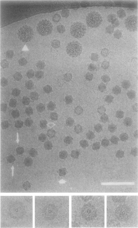
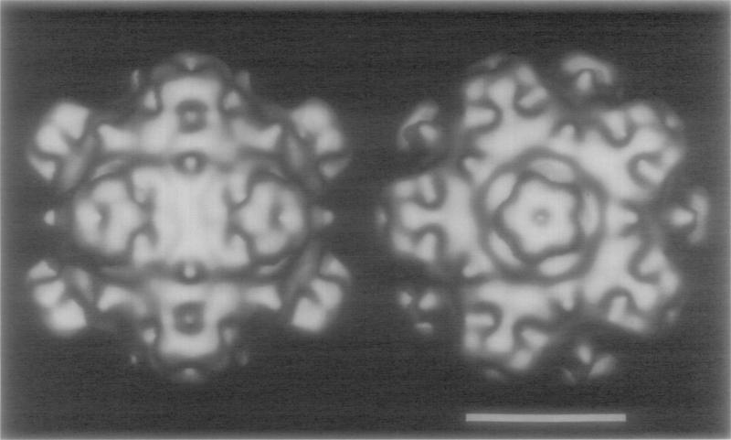
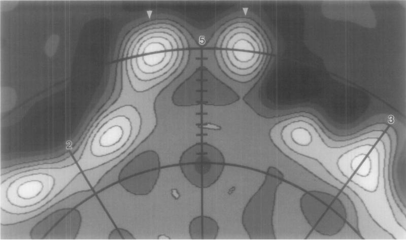
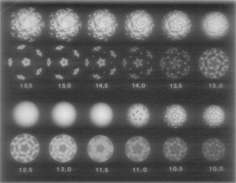
The three-dimensional structure of bacteriophage phi X174 (phi X174) was determined to approximately 2.6 nm resolution from images of frozen-hydrated 114 S particles. The outer surface of phi X174 is characterized by several prominent features: (i) 12 mushroom-shaped caps (approximately 7.1 nm wide x 3.8 nm high) are situated at each of the vertices of the icosahedral virion and extend to a maximum radius of 16.8 nm; (ii) a "collar" of density surrounds the base of each apical cap; and (iii) 20 conical protrusions (approximately 2.3 nm high) lie along the three-fold symmetry axes. The caps have a pentagonal morphology composed of five globular "subunits" and appear to be loosely connected to the underlying capsid. The distribution of the four gene products present in virions (60 copies each of gpF, gpG, and gpJ, and 12 copies of gpH), and the single-stranded DNA (ssDNA) genome cannot be directly discerned in the reconstructed density map, although plausible assignments can be made on the basis of solvent-excluded volume estimates and previous biochemical data. Thus, gpG accounts for most of the mass in the caps; gpH, a presumed cap protein, cannot be identified in part due to the symmetry-averaging procedures, but may be partially located within the interior of the capsid; and gpF and gpJ make up the remainder of the capsid. The genome appears to be less densely packaged inside the capsid compared to many dsDNA viruses whose nucleic acid is arranged in a liquid-crystalline state.
Figures
References
-
- Air GM, Coulson AR, Fiddes JC, Friedmann T, Hutchison CA,, III, Sanger F, Slocombe PM, Smith AJH. J. Mol. Biol. 1978;125:247–254. - PubMed
-
- American Heritage Dictionary, Second College Ed . Houghton Mifflin Co.; Boston, MA.: 1985.
Publication types
MeSH terms
Substances
Grants and funding
LinkOut - more resources
Full Text Sources
Other Literature Sources
Miscellaneous
