. 2004 Feb;3(1):61-71.
doi: 10.1128/EC.3.1.61-71.2004.

The Ca2+/calcineurin-regulated cup gene family in Dictyostelium discoideum and its possible involvement in development

Affiliations

The Ca2+/calcineurin-regulated cup gene family in Dictyostelium discoideum and its possible involvement in development

Barrie Coukell et al. Eukaryot Cell. 2004 Feb.

Abstract

Changes in free intracellular Ca2+ are thought to regulate several major processes during Dictyostelium development, including cell aggregation and cell type-specific gene expression, but the mechanisms involved are unclear. To learn more about Ca2+ signaling and Ca2+ homeostasis in this organism, we used suppression subtractive hybridization to identify genes up-regulated by high extracellular Ca2+. Unexpectedly, many of the genes identified belong to a novel gene family (termed cup) with seven members. In vegetative cells, the cup genes were up-regulated by high Ca2+ but not by other ions or by heat, oxidative, or osmotic stress. cup induction by Ca2+ was blocked completely by inhibitors of calcineurin and protein synthesis. In developing cells, cup expression was high during aggregation and late development but low during the slug stage. This pattern correlates closely with reported levels of free intracellular Ca2+ during development. The cup gene products are highly homologous, acidic proteins possessing putative ricin domains. BLAST searches failed to reveal homologs in other organisms, but Western analyses suggested that Cup-like proteins might exist in certain other cellular slime mold species. Localization experiments indicated that Cup proteins are primarily cytoplasmic but become cell membrane-associated during Ca2+ stress and cell aggregation. When cup expression was down-regulated by antisense RNA, the cells failed to aggregate. However, this developmental block was overcome by partially up-regulating cup expression. Together, these results suggest that the Cup proteins in Dictyostelium might play an important role in stabilizing and/or regulating the cell membrane during Ca2+ stress and/or certain stages of development.


Figures

FIG. 6.
FIG. 6.
Cellular localization of the Cup proteins. (A) Indirect immunofluorescence of AX2 cells either untreated (a) or treated with 50 mM CaCl2 for 4 h (b and c), vegetative CupB-overexpressing (OE) cells (d), or aggregating AX2 cells (e). Scale bar, 5 μm. (B) Cell fractionation. Ca2+-stressed AX2 cells (1.5 × 10⁷/ml) were disrupted by freeze-thaw lysis in the presence of EDTA or CaCl2 and centrifuged, and equal volumes of supernatant (S) and resuspended pellet (P) were analyzed by Western blotting using CupA-N antibodies. Proteins in the incubation medium concentrated ∼100-fold were also analyzed (E).
FIG. 1.
FIG. 1.
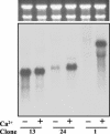
Northern blot analysis of representative clones isolated by SSH. Total RNA (20 μg/lane) from AX2 cells untreated (−) or treated (+) with 80 mM CaCl2 for 3 h was fractionated by gel electrophoresis, stained with ethidium bromide (upper panel), transferred to a nylon membrane, and probed with the cDNA inserts of SSH clone 13, 24, or 1 (lower panel). Radioactivity associated with each band was quantified on an InstantImager.
FIG. 2.
FIG. 2.
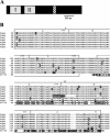
(A) Domain structure of the Cup proteins. Locations of the two ricin domains (I and II) and the coiled-coil region (cross-hatched) in most of the Cup proteins are indicated. (B) Amino acid sequences of the ricin domains in the Cup proteins (CupA to -G) and the hypothetical Dictyostelium protein AAO51776 (51776) (see Discussion). Ricin I and II domains are boxed, and the approximate boundaries of the subdomains (a to c) are indicated. The signature Q-X-W motifs are indicated by asterisks. Amino acids in each protein are shown on the left. For AAO51776, only amino acids in the putative ricin domain are presented. Identical amino acids at each site are shown with white backgrounds, while similar and different amino acids have gray and black backgrounds, respectively.
FIG. 3.
FIG. 3.
Analysis of CupA expression. (A) DNA sequence of the cupA gene in the region of the internal TGA stop codon (base pairs 1270 to 1272). Nucleotides are shown in codons corresponding to ORF-1. *, stop codons in this reading frame. Overlined sequences denote the −1 reading frame, ORF-2. Putative slippery heptameric sequences (see Discussion) preceding the TGA are underlined. (B) Northern blot analysis of total RNA (20 μg/lane) isolated from cupA- and cupB-overexpressing (OE) cells growing axenically in medium containing 20 μg of G418/ml. The membrane was probed with the cDNA insert of SSH clone 1. (C) Western blot analysis of Cup protein expression. Lysates (1.5 × 10⁶ cells/lane) of the cupA- and cupB-overexpressing cells and AX2 cells either untreated (−) or treated (+) with 50 mM CaCl2 for 4 h were fractionated and analyzed as described in Materials and Methods. Molecular mass markers are shown on the left.
FIG. 4.
FIG. 4.
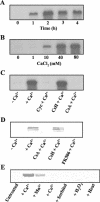
Regulation of cup gene expression in vegetative cells. In all experiments, mid-log-phase AX2 cells were shaken in MES-HL5 medium at 1 × 10⁷ to 2 × 10⁷ cells/ml. (A) Time course of cup up-regulation by Ca2+. Northern blot analysis of total RNA from cells treated with 80 mM CaCl2 for 0, 1, 2, 3, or 4 h. (B) Dose response of cup up-regulation by Ca2+. Northern blot analysis of total RNA from cells treated with 0, 1, 10, 40, or 80 mM CaCl2 for 2 h. (C) Effects of inhibitors of protein synthesis or calcineurin on cup mRNA expression. Northern blot analysis of total RNA from cells either untreated (−) or treated (+) with 80 mM CaCl2 for 2 h in the absence or presence of cycloheximide (Cyc), cyclosporine H (CsH), or cyclosporine A (CsA). Northern membranes were probed with the cDNA insert of SSH clone 1. (D) Effects of inhibitors of calcineurin on Cup protein expression. Western blot analysis of lysates of cells (8 × 10⁵/lane) either untreated (−) or treated (+) with 50 mM CaCl2 for 4 h in the absence or presence of cyclosporine A (CsA), cyclosporine H (CsH), or FK506. (E) Effects of various environmental stressors on Cup protein expression. Western blot analysis of lysates of cells (1.5 × 10⁶/lane) either untreated or treated with different stressors for 3 h (see Materials and Methods for conditions). Western blots were probed with the CupA-N antibodies.
FIG. 5.
FIG. 5.
Regulation of cup gene expression during development. (A) Northern blot analysis of cup expression in cells developing on PBS agar. Nonaxenically grown AX2 cells were washed free of bacteria and plated on PBS agar at a density of 3.2 × 10⁶ cells/cm2.The plates were incubated in a humid environment at 22°C, and total RNA was isolated from cells at 3-h intervals. Developmental timing: 0 h, vegetative cells; 6 to 9 h, aggregation; 12 h, tipped aggregates; 15 h, slugs; 18 to 21 h, culmination; 24 h, fruiting bodies. (B) Effect of Ca2+ on cup expression during development. Nonaxenically grown AX2 cells were washed free of bacteria in 20 mM MES-NaOH (pH 6.6) and plated at the same density as in panel A on agar prepared in the same MES buffer with or without 20 mM CaCl2. At the times indicated, total RNA was isolated from cells and used in Northern blot analysis. The developmental timing under these conditions was approximately the same as on PBS agar. Northern blots were probed with the cDNA insert from SSH clone 1.
FIG. 7.
FIG. 7.
Effect of antisense cupB RNA expression on Dictyostelium development. (A) Western blot analysis of lysates (1.5 × 10⁶ cells/lane) of control (vector alone) and cupB antisense (AS) cells either untreated (−) or treated (+) with 50 mM CaCl2 for 4 h. (B) Development of control and cupB antisense cells. Mid-log-phase control (a and c) and antisense (b and d) cells were harvested, washed in 17 mM phosphate, plated (20 μl of 2 × 10⁸ cells/ml) on MES agar either without (a and b) or with (c and d) 20 mM CaCl2, and incubated in a humid environment for 24 h. Scale bar, 0.5 mm.
FIG. 8.
FIG. 8.
Cup-like proteins in other cellular slime mold species. Cells of D. discoideum (D.d.), P. violaceum (P.v.), A. leptosomum (A.l.), and D. lacteum (D.l.) were grown as described in Materials and Methods, washed, and shaken in MES-HL5 medium at 1 × 10⁷ cells/ml for 4 h either without (−) or with (+) 50 mM CaCl2. Lysates of D. discoideum cells (8 × 10⁵/lane) and other cells (5 × 10⁶/lane) were analyzed by Western blotting using CupA-N antibodies.

Similar articles

Cited by

References

    1. Abe, T., and Y. Maeda. 1989. The prestalk/prespore differentiation and polarized cell movement in Dictyostelium discoideum slugs. A possible involvement of the intracellular Ca2+ concentration. Protoplasma 151:175-178.
    1. Aichem, A., and R. Mutzel. 2001. Unconventional mRNA processing in the expression of two calcineurin B isoforms in Dictyostelium. J. Mol. Biol. 308:873-882. - PubMed
    1. Baskar, R., P. Chhabra, P. Mascarenhas, and V. Nanjundiah. 2000. A cell type-specific effect of calcium on pattern formation and differentiation in Dictyostelium discoideum. Ind. J. Dev. Biol. 44:491-498. - PubMed
    1. Blumberg, D., J. F. Comer, and E. M. Walton. 1989. Ca++ antagonists distinguish different requirements for cAMP-mediated gene expression in the cellular slime mold, Dictyostelium discoideum. Differentiation 41:14-21. - PubMed
    1. Bonner, J. T., E. M. Hall, S. Noller, F. B. Oleson, Jr., and A. B. Roberts. 1972. Synthesis of cyclic AMP and phosphodiesterase in various species of cellular slime molds and its bearing on chemotaxis and differentiation. Dev. Biol. 29:402-409. - PubMed

Publication types

MeSH terms