

Higher atrophy rate of entorhinal cortex than hippocampus in AD
- PMID: 14872024
- PMCID: PMC1820859
- DOI: 10.1212/01.wnl.0000106462.72282.90
Higher atrophy rate of entorhinal cortex than hippocampus in AD
Abstract
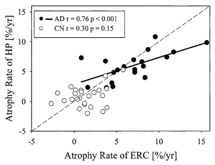
Objectives: To determine if atrophy rates were higher for entorhinal cortex (ERC) than for hippocampus in Alzheimer disease (AD), to determine the relationship between hippocampal atrophy rate and memory impairment, and to compare atrophy rates of ERC and hippocampus in differentiating between patients with AD and cognitively normal (CN) controls.
Methods: Twenty patients with AD and 25 CN subjects had MRI scans and clinical evaluations twice approximately 1.9 years apart. ERC volumes were measured manually and hippocampal volumes were measured semiautomatically on volumetric T1-weighted MR images.
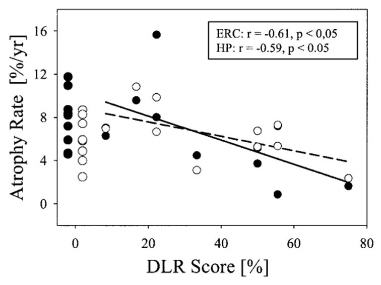
Results: In AD, the atrophy rate of ERC (7.1 +/- 3.2%/year) was higher (p < 0.02) than that of hippocampus (5.9 +/- 2.4%/year). Furthermore, memory deficit in mild AD, measured with the Delayed List Verbal Recall test, correlated significantly with atrophy rates of both ERC (r = -0.61) and hippocampus (r = -0.59). Atrophy rates of ERC and hippocampus were comparable in differentiating between AD and CN. Using atrophy rates of ERC or hippocampus to detect a 20% treatment effect with 90% power (p < 0.05) would require about 100 completed patients per arm in a 2-year study.
Conclusion: The finding in AD that the atrophy rate in the entorhinal cortex is higher than in the hippocampus is consistent with the view that AD pathology begins in the entorhinal cortex.
Figures
Similar articles
-
Atrophy rates of entorhinal cortex in AD and normal aging.Neurology. 2003 Feb 11;60(3):481-6. doi: 10.1212/01.wnl.0000044400.11317.ec. Neurology. 2003. PMID: 12578931 Free PMC article. Clinical Trial.
-
Magnetic resonance imaging of the entorhinal cortex and hippocampus in mild cognitive impairment and Alzheimer's disease.J Neurol Neurosurg Psychiatry. 2001 Oct;71(4):441-7. doi: 10.1136/jnnp.71.4.441. J Neurol Neurosurg Psychiatry. 2001. PMID: 11561025 Free PMC article.
-
Effects of subcortical ischemic vascular dementia and AD on entorhinal cortex and hippocampus.Neurology. 2002 Jun 11;58(11):1635-41. doi: 10.1212/wnl.58.11.1635. Neurology. 2002. PMID: 12058091 Free PMC article.
-
Neuroimaging of hippocampal atrophy in early recognition of Alzheimer's disease--a critical appraisal after two decades of research.Psychiatry Res Neuroimaging. 2016 Jan 30;247:71-8. doi: 10.1016/j.pscychresns.2015.08.014. Psychiatry Res Neuroimaging. 2016. PMID: 26774855 Review.
-
From healthy aging to early Alzheimer's disease: in vivo detection of entorhinal cortex atrophy.Ann N Y Acad Sci. 2000 Jun;911:240-53. doi: 10.1111/j.1749-6632.2000.tb06730.x. Ann N Y Acad Sci. 2000. PMID: 10911878 Review.
Cited by
-
Selective interaction of lansoprazole and astemizole with tau polymers: potential new clinical use in diagnosis of Alzheimer's disease.J Alzheimers Dis. 2010;19(2):573-89. doi: 10.3233/JAD-2010-1262. J Alzheimers Dis. 2010. PMID: 20110603 Free PMC article.
-
A multivariate model of time to conversion from mild cognitive impairment to Alzheimer's disease.Geroscience. 2020 Dec;42(6):1715-1732. doi: 10.1007/s11357-020-00260-7. Epub 2020 Sep 4. Geroscience. 2020. PMID: 32886293 Free PMC article.
-
Differential Gray Matter Vulnerability in the 1 Year Following a Clinically Isolated Syndrome.Front Neurol. 2018 Oct 11;9:824. doi: 10.3389/fneur.2018.00824. eCollection 2018. Front Neurol. 2018. PMID: 30364223 Free PMC article.
-
Comparisons between global and focal brain atrophy rates in normal aging and Alzheimer disease: Boundary Shift Integral versus tracing of the entorhinal cortex and hippocampus.Alzheimer Dis Assoc Disord. 2004 Oct-Dec;18(4):196-201. Alzheimer Dis Assoc Disord. 2004. PMID: 15592130 Free PMC article.
-
Influence of MRI on Diagnostic Efficacy and Satisfaction of Patients with Alzheimer's Disease.Comput Math Methods Med. 2021 Nov 8;2021:9038784. doi: 10.1155/2021/9038784. eCollection 2021. Comput Math Methods Med. 2021. Retraction in: Comput Math Methods Med. 2022 Dec 27;2022:9761394. doi: 10.1155/2022/9761394. PMID: 34790255 Free PMC article. Retracted.
References
-
- Squire LR, Zola-Morgan S. The medial temporal lobe memory system. Science. 1991;253:1380–1386. - PubMed
-
- Braak H, Braak E. Neuropathological stageing of Alzheimer-related changes. Acta Neuropathol (Berl) 1991;82:239–259. - PubMed
-
- Haroutunian V, Purohit DP, Perl DP, et al. Neurofibrillary tangles in nondemented elderly subjects and mild Alzheimer disease. Arch Neurol. 1999;56:713–718. - PubMed
-
- Haroutunian V, Perl DP, Purohit DP, et al. Regional distribution of neuritic plaques in the nondemented elderly and subjects with very mild Alzheimer disease. Arch Neurol. 1998;55:1185–1191. - PubMed
-
- Bobinski M, de Leon MJ, Convit A, et al. MRI of entorhinal cortex in mild Alzheimer’s disease. Lancet. 1999;353:38–40. - PubMed
Publication types
MeSH terms
Grants and funding
LinkOut - more resources
Full Text Sources
Other Literature Sources
Medical