. 2004 Feb 10;32(3):997-1005.
doi: 10.1093/nar/gkh265. Print 2004.

Phosphatidyl inositol 3-kinase-like serine/threonine protein kinases (PIKKs) are required for DNA damage-induced phosphorylation of the 32 kDa subunit of replication protein A at threonine 21

Affiliations

Phosphatidyl inositol 3-kinase-like serine/threonine protein kinases (PIKKs) are required for DNA damage-induced phosphorylation of the 32 kDa subunit of replication protein A at threonine 21

Wesley D Block et al. Nucleic Acids Res. .

Abstract

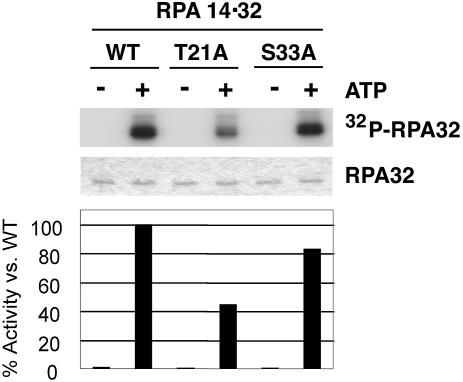
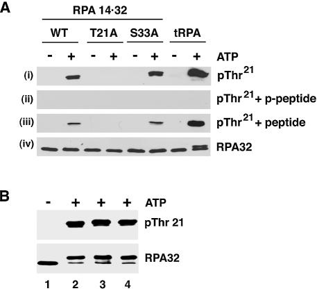
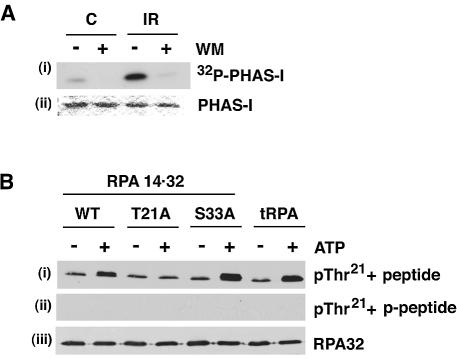
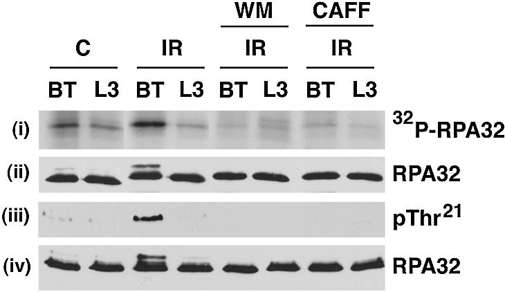
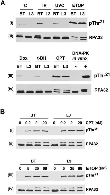
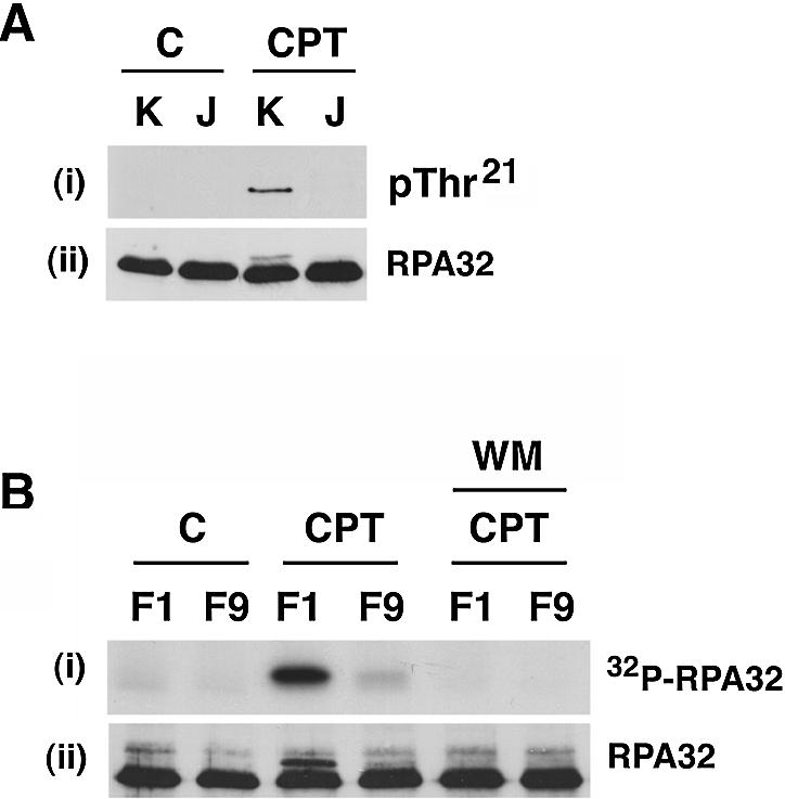
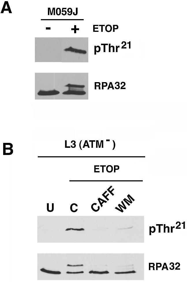
Replication protein A (RPA) is a single-stranded DNA (ssDNA) binding protein involved in various processes, including nucleotide excision repair and DNA replication. The 32 kDa subunit of RPA (RPA32) is phosphorylated in response to various DNA-damaging agents, and two protein kinases, ataxia-telangiectasia mutated (ATM) and the DNA-dependent protein kinase (DNA-PK) have been implicated in DNA damage-induced phosphorylation of RPA32. However, the relative roles of ATM and DNA-PK in the site-specific DNA damage-induced phosphorylation of RPA32 have not been reported. Here we generated a phosphospecific antibody that recognizes Thr21-phosphorylated RPA32. We show that both DNA-PK and ATM phosphorylate RPA32 on Thr21 in vitro. Ionizing radiation (IR)-induced phosphorylation of RPA32 on Thr21 was defective in ATM-deficient cells, while camptothecin (CPT)-induced phosphorylation of RPA32 on Thr21 was defective in cells lacking functional DNA-PK. Neither ATM nor DNA-PK was required for etoposide (ETOP)-induced RPA32 Thr21 phosphorylation. However, two inhibitors of the ATM- and Rad3-related (ATR) protein kinase activity prevented ETOP-induced Thr21 phosphorylation. Inhibition of DNA replication prevented both the IR- and CPT-induced phosphorylation of Thr21, whereas ETOP-induced Thr21 phosphorylation did not require active DNA replication. Thus, the regulation of RPA32 Thr21 phosphorylation by multiple DNA damage response protein kinases suggests that Thr21 phosphorylation of RPA32 is a crucial step within the DNA damage response.


Figures

Figure 1
Figure 1
Thr21 is the major in vitro DNA-PK phosphorylation site in RPA32. One microgram of WT-, T21A- or S33A-RPA14·32 was incubated with purified DNA-PK (60 ng of DNA-PKcs and 20 ng of Ku70/80) in the presence or absence of [γ-32P]ATP for 10 min. Samples were fractionated on SDS–polyacrylamide gels and RPA32 was visualized by Coomassie staining (RPA32) and autoradiography (32P-RPA32). RPA32 bands were excised from the gel and 32P incorporation was quantitated by Cerenkov counting (bottom).
Figure 2
Figure 2
In vitro phosphorylation of RPA32 by DNA-PK. (A) Seventy-two nanograms of WT-, T21A- or S33A-RPA14·32, or 100 ng of tRPA were incubated with purified DNA-PK (as in Fig. 1) in the presence or absence of ATP. Samples were immunoblotted using the Thr21 phosphospecific antibody (pThr21) either alone (i), in the presence of pThr21 blocking peptide (p-peptide) (ii), or in the presence of Thr21 mock peptide (peptide) (iii). Alternatively, samples were immunoblotted using a monoclonal RPA32 antibody (RPA32) (iv). (B) tRPA (100 ng) was incubated with purified DNA-PK in the presence or absence of ATP for 5 min, with additional DNA added to the incubation as follows: lanes 1 and 2, none; lane 3, M13 closed circular ssDNA; lane 4, 40 nt ssDNA. Samples were immunoblotted using the Thr21 phosphospecific antibody (pThr21) or a monoclonal RPA32 antibody (RPA32).
Figure 3
Figure 3
In vitro phosphorylation of RPA32 by ATM. (A) ATM was immunoprecipitated from 1 mg of protein extract from unirradiated ‘C’ cells or cells exposed to 10 Gy IR as described in Materials and Methods. Immunoprecipitates were assayed in the presence of [γ-32P]ATP and 0.5 µg of PHAS-I, in the presence or absence of 1 µM wortmannin (WM). PHAS-I was visualized by Coomassie staining (PHAS-I) or autoradiography (32P-PHAS-I). (B) ATM immunoprecipitates from irradiated cells were incubated with 0.75 µg of WT, T21A or S33A RPA14·32, or 1 µg of tRPA, in the presence or absence of 250 µM ATP for 30 min. Samples were immunoblotted using the Thr21 phosphospecific antibody (pThr21) either in the presence of Thr21 mock peptide (peptide) (i), or in the presence of pThr21 blocking peptide (p-peptide) (ii). Alternatively, samples were immunoblotted using a monoclonal RPA32 antibody (RPA32) (iii).
Figure 4
Figure 4
IR-induced RPA phosphorylation is ATM dependent. (i and ii) BT (ATM-proficient) or L3 (ATM-deficient) cells were labeled with [32P]orthophosphate, exposed to 10 Gy IR or unirradiated ‘C’, with either 100 µM wortmannin (WM) or 10 mM caffeine (CAFF) pre-treatment as described in Materials and Methods. RPA32 was immunoprecipitated from cell extracts and phosphorylated RPA32 was visualized by autoradiography [32P-RPA32 (i)] or with an RPA32 antibody [RPA32 (ii)]. (iii and iv) Cells were treated as in (i) and (ii) but without [32P]orthophosphate. Cell extracts were enriched for RPA32 using Affi-gel blue resin as described in Materials and Methods, and RPA32 was immunoblotted using the Thr21 phosphospecific antibody [pThr21 (iii)] or a RPA32 antibody [RPA32 (iv)].
Figure 5
Figure 5
ATM-dependent and -independent DNA damage-induced phosphorylation of RPA32 Thr21. (A) BT (ATM-proficient) or L3 (ATM- deficient) cells were treated with the indicated DNA-damaging agent as described in Materials and Methods. Cell extracts were enriched for RPA32 using Affi-gel blue resin and RPA32 was immunoblotted using the Thr21 phosphospecific antibody (pThr21) or a RPA32 antibody (RPA32). C, IR, UVC, ETOP, Dox, t-BH and CPT denote control (untreated), ionizing radiation (10 Gy), ultraviolet radiation (254 nm; 20 J/m2), etoposide (68 µM), doxorubicin (10 µM), t-butyl hydroperoxide (500 µM), and camptothecin (100 µM), respectively. (B) BT (ATM-proficient) or L3 (ATM-deficient) cells were treated with a range of CPT or ETOP concentrations and processed as in (A).
Figure 6
Figure 6
Involvement of DNA replication in the DNA damage-induced phosphorylation of RPA32 Thr21. (A) BT (ATM-proficient) cells were either exposed to 10 Gy IR or left untreated ‘C’, either with or without 1 h pre-treatment with aphidicolin. Cell extracts were enriched for RPA32 using Affi-gel blue resin and RPA32 was immunoblotted using the Thr21 phosphospecific antibody (pThr21) or a RPA32 antibody (RPA32). (B) L3 (ATM-deficient) cells were treated with 10 µM CPT, 68 µM ETOP or were left untreated ‘C’, either with or without 1 h pre-treatment with aphidicolin, and processed as in (A).
Figure 7
Figure 7
DNA-PK regulates the CPT-induced RPA32 Thr21 phosphorylation. (A) DNA-PKcs-proficient M059K (K) and DNA-PKcs-deficient M059J (J) cells were untreated ‘C’ or treated with 10 µM CPT and analyzed for RPA32 Thr21 phosphorylation, as in Figure 5A. (B) M059J/Fus1 (DNA-PKcs-proficient) and M059J/Fus9 (DNA-PKcs-deficient) cells were labeled with [32P]orthophosphate and were either left without additional treatment ‘C’ or treated with 10 µM CPT, with or without pre-treatment with 100 µM wortmannin. RPA32 was immunoprecipitated from cell extracts and visualized by autoradiography (32P-RPA32) or with an RPA32 antibody (RPA32).
Figure 8
Figure 8
DNA-PK-independent ETOP-induced RPA32 Thr21 phosphorylation. (A) DNA-PKcs-deficient M059J cells were treated with ETOP and analyzed for RPA32 Thr21 phosphorylation, as in Figure 5A. (B) L3 (ATM-deficient) cells were untreated ‘U’ or treated with 68 µM ETOP, either with ETOP alone ‘C’ or with pre-treatment with 10 mM CAFF or 100 µM wortmannin, and then analyzed for RPA32 Thr21 phosphorylation, as in Figure 5A.

References

    1. Wold M.S. (1997) Replication protein A: a heterotrimeric, single-stranded DNA-binding protein required for eukaryotic DNA metabolism. Annu. Rev. Biochem., 66, 61–92. - PubMed
    1. Dutta A. and Stillman,B. (1992) cdc2 family kinases phosphorylate a human cell DNA replication factor, RPA and activate DNA replication. EMBO J., 11, 2189–2199. - PMC - PubMed
    1. Din S., Brill,S.J., Fairman,M.P. and Stillman,B. (1990) Cell-cycle-regulated phosphorylation of DNA replication factor A from human and yeast cells. Genes Dev., 4, 968–977. - PubMed
    1. Liu V.F. and Weaver,D.T. (1993) The ionizing radiation-induced replication protein A phosphorylation response differs between ataxia telangiectasia and normal human cells. Mol. Cell. Biol., 13, 7222–7231. - PMC - PubMed
    1. Brush G.S., Morrow,D.M., Hieter,P. and Kelly,T.J. (1996) The ATM homologue MEC1 is required for phosphorylation of replication protein A in yeast. Proc. Natl Acad. Sci. USA, 93, 15075–15080. - PMC - PubMed

Publication types

MeSH terms