. 1951 Nov;14(6):479-96.
doi: 10.1152/jn.1951.14.6.479.

Collateral afferent excitation of reticular formation of brain stem

Collateral afferent excitation of reticular formation of brain stem

T E STARZL et al. J Neurophysiol. 1951 Nov.
No abstract available


Figures

Fig. 1
Fig. 1
Transverse sections through hemisphere with shading indicating areas from which evoked potentials are recorded upon single-shock stimulation of sciatic nerve. Cross-hatched zones are regions of greatest response potentiality, and are seen to be in medial brain stem areas as well as along through sensory path. Only B-erythroidine preparations included. Abbreviations for Figs. 1 and 5 are as follows: A—amygdala, AM—anteromedial nuc., AV—anteroventral nuc., BIC—brachium inferior colliculus, BP—basis pedunculi, C—caudate nuc., CE—nuc. centralis medialis, CG—central gray, CL—nuc. centralis lateralis, CM—centre median, F—fornix, GMES—middle ectosylvian gyrus, GP—globus pallidus, H—habenular nuclei, HP—habenulo-peduncular tract, HVM—ventromedial nuc. of hypothalamus, IAM—intermediate anteromedial nuc., LA—nuc. lateral anterior, LG—lateral geniculate nuc., LP—nuc. lateralis posterior, M—medial nuc., MB—mammillary body, MG—medial geniculate nuc., ML—medial lemniscus, NR—red nuc., OT—optic tract, P—posterior nuc., PL—pulvinar, PRE—pretectal region, PT—putamen, RE—nuc. reuniens, RT—reticular nuc., SC—superior colliculus, SN—substantia nigra, SU—subthalamic nuc., VA—nuc. ventralis anterior, VL—nuc. ventralis lateralis, VM—ventromedial nuc, VPL—ventroposterolateral nuc., VPM—ventroposteromedial nuc.
Fig. 2
Fig. 2
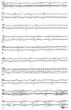
Records of collateral afferent somatic potentials evoked by stimulation of left sciatic nerve with 3–5 volts. Responses illustrated are from red nucleus (A), tegmentum of midbrain (B), pretectal region (C), subthalamus (D), ventromedial thalamic nucleus (E), ventralis lateralis (F), lateralis posterior (G), reuniens (H), ventromedial to centralis lateralis (I), and ventralis anterior (J). All deep pickups are on right side, contralateral to stimulus, except J which is ipsilaterally located. In each case, activity of right sensory cortex (R SEN) is recorded in upper channel. Notice waxing and waning of evoked potentials in G, and tripping of spindles in I and J. Individual stimuli are marked with arrows. All under B-erythroidine.
Fig. 3
Fig. 3
Records illustrating various characteristics of collateral somatic afferent potentials upon stimulation of sciatic nerve with 3–5 volts. In A is shown phase reversal of response on passing through centre median-subthalamic region, electrode having been lowered by 1 mm. steps (1–6). In B is illustrated the differing effects, in both subthalamic and cortical leads, with different frequencies of sciatic stimulation. Frequencies used were 6/sec. (i), 15/sec. (2), 25/sec. (5), and 50/sec. (4). In C are seen the large collateral potentials in red nucleus area with sciatic stimulation at 3/sec. (1), or 10/sec. (2). Notice increase in background activity produced by stimulation and its persistence as an after-discharge in both cases. D illustrates effects of frequency variation on collateral potentials recorded from midbrain tegmentum; sciatic nerve was stimulated (from left to right) at 10, 20, 30, and 50/sec. Deep pickups are on side of sciatic stimulation in A and B, and contralateral in C and D. In each strip, upper channel records the activity of sensory cortex contralateral to side of stimulus, except B which is ipsilateral. Arrows indicate single stimuli, and dark lines mark periods of repetitive excitation. Cortical abbreviations are: SEN—sensory, SEN-MOT—sensory motor. All under B-erythroidine.
Fig. 4
Fig. 4
Records showing collateral somatic afferent potentials evoked by sciatic stimulation (3–5 volts), after various lesions. After complete cerebellectomy plus bilateral extirpation of somatic cortex (A), responses are evoked by sciatic stimulation in region of ventromedial nucleus and subthalamus (1), centre median (2), superior colliculus (3), and from red nucleus (4). In recording between subthalamus and ventromedial nucleus, the full response is seen with stimuli at 1.2/sec. (A, 1—left), but with 3/sec, the wave component falls out leaving a spike (1—right). After lesions of medial midbrain tegmentum (B), collateral responses are still found at more rostral levels. In B, 1, such a medial lesion was placed at intercollicular level leaving only lateral sensory pathways intact, and collateral potentials were recorded between centralis lateralis and ventromedial nuclei of thalamus. In B, 2 and 3, a lesion was placed at tegmento-thalamic junction, sparing only laterally located sensory pathways. Electrodes placed in centre median (B, 2), and between subthalamus and ventromedial nucleus (B, 3), revealed continued presence of medially located collateral potentials. Complete bilateral destruction of medial lemnisci at intercollicular level, sparing medial tegmentum, does not abolish collateral responses at more rostral levels (C) as shown from ventromedial nucleus (C, 1), and upper midbrain tegmentum (C, 2). With lemnisci eliminated, sensory cortex does not exhibit a primary spike, but sciatic stimulation still appears to have an initiating effect on bursts (C, 1). Subcortical pickups were ipsilateral to side of sciatic stimulation (A, 1–3; B, 1–2), and contralateral (A, 4; B, 3; C, 1–2). All sensory cortical pickups (R. SEN) were contralateral to stimulus. Shocks are marked by arrows. All under B-erythroidine.
Fig. 5
Fig. 5
Transverse sections through hemisphere, with shading indicating areas from which evoked potentials are recorded upon click stimulation. Cross-shading areas indicate regions of best response. As with somatic system, large medial area of brain stem is implicated, as well as through auditory pathway. Only B-erythroidine preparations included. In D and E, lateral part of medial geniculate was not explored.
Fig. 6
Fig. 6
Records of collateral auditory potentials, evoked by click stimuli, in midbrain tegmentum (A), superior colliculus (B), red nucleus (C), centre median (D, E), subthalamus (F), lateral hypothalamus (G), globus pallidus at its junction with putamen (H), and ventralis anterior nucleus (I). Note occasional tripping of spindles in centre median (E) and regular tripping in ventralis anterior (I). Record from auditory cortex (AUD), on same side as deep pickup, is shown in upper channel in each case. Click stimuli are marked by arrows. All under B-erythroidine except I, from an encephale isolé.
Fig. 7
Fig. 7
Records of collateral auditory potentials after various lesions. After bilateral removal of auditory cortex, responses are shown in region of centralis medialis and ventromedial nuclei (A) and midbrain tegmentum (B). After complete bilateral destruction of medial geniculate bodies, collateral potentials are seen in subthalamus and red nucleus (C), former in this case being a broad wave. With complete bilateral destruction of inferior colliculi, responses are shown in subthalamus (D). After complete cerebellectomy, collateral potentials are illustrated in ventromedial thalamic nucleus (E) and midbrain tegmentum (F). After destruction of medial midbrain tegmentum at level of superior colliculus, leaving only lateral sensory pathways intact, collateral auditory responses are shown in subthalamus (G) and red nucleus (H). With such a medial lesion at tegmento-thalamic junction, sparing only lateral sensory paths, a collateral auditory response is seen in ventromedial nucleus (I). Click stimuli are marked with arrows. Abbreviations for cortical leads are: AUD—auditory, MOT—motor. All under B-erythroidine.
Fig. 8
Fig. 8
Outline of brain of cat, showing distribution of afferent collaterals to ascending reticular activating system in brain stem.

References

    1. Ades HW. Midbrain auditory mechanisms in cats. J. Neurophysiol. 1944;7:415–424.
    1. Allen WR. Origin and destination of the secondary visceral fibers in the guinea-pig. J. comp. Neurol. 1923;35:275–311.
    1. Barnes WT, Magoun HW, Ranson SW. The ascending auditory pathway in the brain stem of the monkey. J. comp. Neurol. 1943;79:129–152.
    1. Berry CM, Karl RC, Hinsey JC. Course of spinothalamic and medial lemniscus pathways in cat and Rhesus monkey. J. Neurophysiol. 1950;13:149–156.
    1. Dempsey EW, Morison RS, Morison BR. Some afferent diencephalic pathways related to cortical potentials in the cat. Amer. J. Physiol. 1941;131:718–731.

LinkOut - more resources