

PDX1 homeobox protein expression in pseudopyloric glands and gastric carcinomas
- PMID: 14960508
- PMCID: PMC1773956
- DOI: 10.1136/gut.2003.026609
PDX1 homeobox protein expression in pseudopyloric glands and gastric carcinomas
Abstract
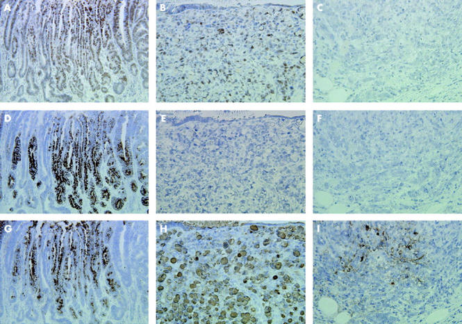
Background and aims: Although it has been reported that intestinal metaplasia implicated in gastric carcinogenesis is induced by the ParaHox gene CDX2, it is unclear which genes are responsible for the formation of pseudopyloric glands and whether they play a role in gastric carcinogenesis. Pancreatic-duodenal homeobox 1 (PDX1) is also a ParaHox gene which contributes to the genesis and development of the pancreas, duodenum, and antrum. To clarify its significance for the formation of pseudopyloric glands and gastric carcinogenesis, we investigated expression of PDX1 and mucin in gastric carcinomas and surrounding mucosa.
Methods: Gastric carcinoma tissues from 95 patients were used for immunohistochemical analyses of PDX1, and mucins MUC6 and MUC5AC.
Results: PDX1 was found to be frequently expressed in pseudopyloric glands and intestinal metaplasia. MUC6 was more abundant than MUC5AC in pseudopyloric glands while higher levels of MUC5AC than MUC6 were evident in intestinal metaplasia. The frequency of PDX1 positive reactivity was higher in differentiated type carcinomas (39/43, 90.7%) and T1 carcinomas (42/43, 97.7%) than in undifferentiated type (33/52, 63.5%) and T2-4 (30/52, 57.7%) carcinomas. PDX1 and MUC6 double positive expression was observed in carcinomas, respectively, including the corpus, and also correlated with histological type and depth of invasion. In contrast, no link was apparent between PDX1 and MUC5AC double positive reactivity and histological type.
Conclusion: Our study suggests that PDX1 plays an important role in the development of pseudopyloric glands, and that pseudopyloric glands may reflect a condition associated with gastric carcinogenesis.
Figures
Similar articles
-
The relationship among PDX1, CDX2, and mucin profiles in gastric carcinomas; correlations with clinicopathologic parameters.J Cancer Res Clin Oncol. 2011 Dec;137(12):1749-62. doi: 10.1007/s00432-011-1044-7. Epub 2011 Sep 10. J Cancer Res Clin Oncol. 2011. PMID: 21909647 Free PMC article.
-
Intestinal metaplasia of human stomach displays distinct patterns of mucin (MUC1, MUC2, MUC5AC, and MUC6) expression.Cancer Res. 1999 Mar 1;59(5):1003-7. Cancer Res. 1999. PMID: 10070955
-
Expression of intestine-specific transcription factors, CDX1 and CDX2, in intestinal metaplasia and gastric carcinomas.J Pathol. 2003 Jan;199(1):36-40. doi: 10.1002/path.1246. J Pathol. 2003. PMID: 12474224
-
Stem cells and gastric cancer: role of gastric and intestinal mixed intestinal metaplasia.Cancer Sci. 2003 Feb;94(2):135-41. doi: 10.1111/j.1349-7006.2003.tb01409.x. Cancer Sci. 2003. PMID: 12708487 Free PMC article. Review.
-
Diagnosis: gastric intestinal metaplasia - what to do next?Curr Opin Gastroenterol. 2019 Nov;35(6):535-543. doi: 10.1097/MOG.0000000000000576. Curr Opin Gastroenterol. 2019. PMID: 31415250 Free PMC article. Review.
Cited by
-
Altered expression of CDX-2, PDX-1 and mucin core proteins in "Ulcer-associated cell lineage (UACL)" in Crohn's disease.J Mol Histol. 2008 Apr;39(2):161-8. doi: 10.1007/s10735-007-9149-7. Epub 2007 Oct 24. J Mol Histol. 2008. PMID: 17957487
-
Negative regulation of pancreatic and duodenal homeobox-1 by somatostatin receptor subtype 5.Mol Endocrinol. 2012 Jul;26(7):1225-34. doi: 10.1210/me.2012-1095. Epub 2012 Jun 5. Mol Endocrinol. 2012. PMID: 22669743 Free PMC article.
-
Tissue MicroArray analyses of pancreatic duodenal homeobox-1 in human cancers.World J Surg. 2005 Mar;29(3):334-8. doi: 10.1007/s00268-004-7823-4. World J Surg. 2005. PMID: 15706433
-
Transcription factor SOX2 up-regulates stomach-specific pepsinogen A gene expression.J Cancer Res Clin Oncol. 2007 Apr;133(4):263-9. doi: 10.1007/s00432-006-0165-x. Epub 2006 Nov 28. J Cancer Res Clin Oncol. 2007. PMID: 17136346 Free PMC article.
-
Mucins, trefoil factors and pancreatic duodenal homeobox 1 expression in spasmolytic polypeptide expressing metaplasia and intestinal metaplasia adjacent to gastric carcinomas.Arch Med Sci. 2013 Aug 12;16(6):1402-1410. doi: 10.5114/aoms.2013.36923. eCollection 2020. Arch Med Sci. 2013. PMID: 33224340 Free PMC article.
References
-
- Laurén P. The two histological main types of gastric carcinoma. Acta Pathol Microbiol Scand 1965;64:34–49. - PubMed
-
- Fenoglio-Preiser C, Muñoz N, Carneiro F, et al. Gastric carcinoma. In: Hamilton SR, Aaltonen LA, eds. World Health Organization classification of tumours. Pathology and genetics of tumours of the digestive system. Lyon: IARC Press, 2000:39–52.
-
- Japanese Gastric Cancer Association. Japanese classification of gastric carcinoma. 2nd English edition. Gastric Cancer 1998;1:10–24. - PubMed
-
- Lewin KJ, Appelman HD. Carcinoma of the stomach. Atlas of tumor pathology, tumors of the esophagus and stomach, 3rd edn. Washington DC: Armed Forces Institute of Pathology, 1995:245–330.
-
- Nakamura K, Sugano H, Takagi K. Carcinoma of the stomach in incipient phase: its histogenesis and histological appearances. Gann 1968;59:251–8. - PubMed
Publication types
MeSH terms
Substances
LinkOut - more resources
Full Text Sources
Other Literature Sources
Medical
Research Materials