

Enteric dopaminergic neurons: definition, developmental lineage, and effects of extrinsic denervation
- PMID: 14960604
- PMCID: PMC6730344
- DOI: 10.1523/JNEUROSCI.3982-03.2004
Enteric dopaminergic neurons: definition, developmental lineage, and effects of extrinsic denervation
Abstract
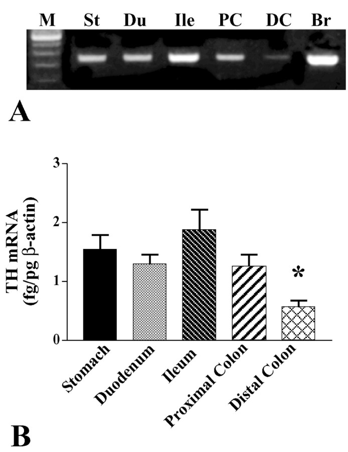
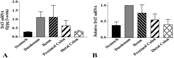
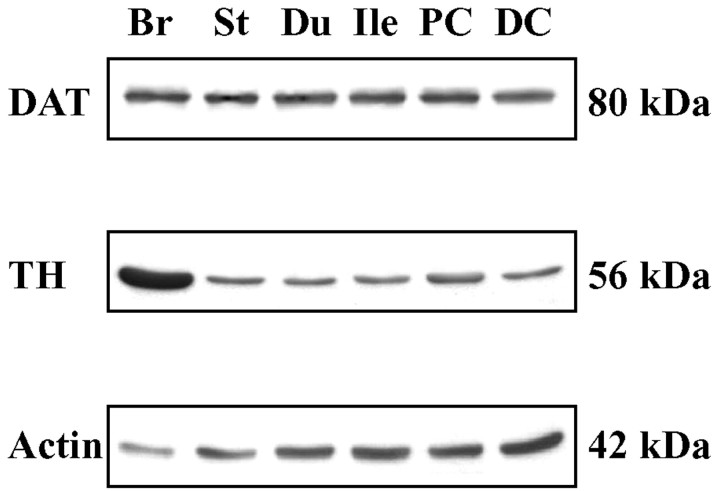
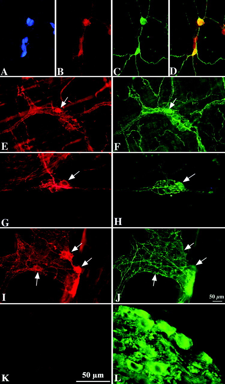
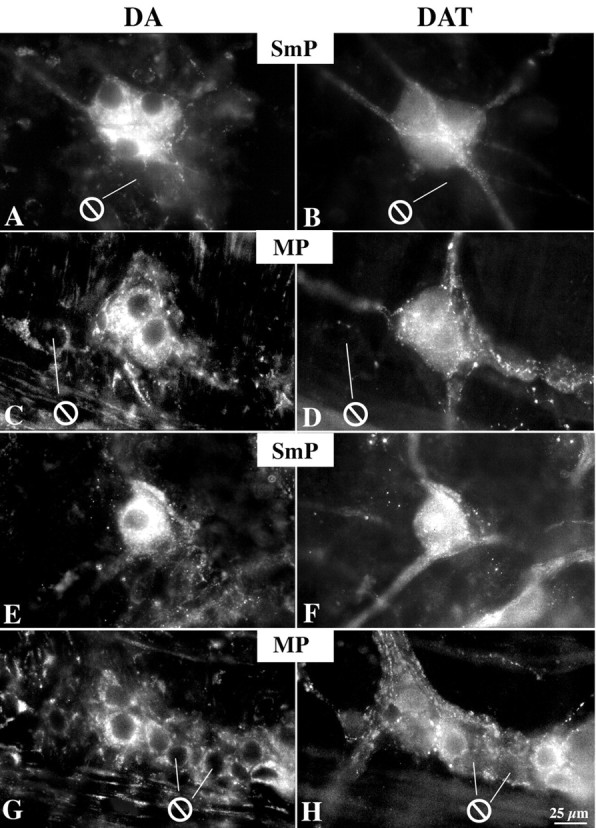
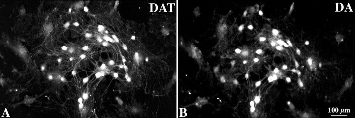
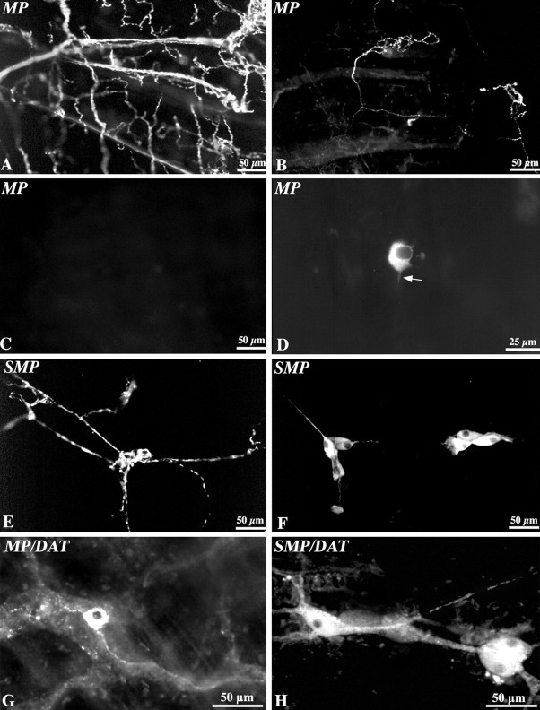
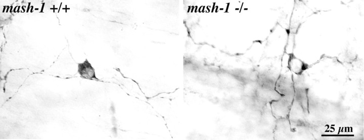
The existence of enteric dopaminergic neurons has been suspected; however, the innervation of the gut by sympathetic nerves, in which dopamine (DA) is the norepinephrine precursor, complicates analyses of enteric DA. We now report that transcripts encoding tyrosine hydroxylase (TH) and the DA transporter (DAT) are present in the murine bowel (small intestine > stomach or colon; proximal colon > distal colon). Because sympathetic neurons are extrinsic, transcripts encoding TH and DAT in the bowel are probably derived from intrinsic neurons. TH protein was demonstrated immunocytochemically in neuronal perikarya (submucosal >> myenteric plexus; small intestine > stomach or colon). TH, DA, and DAT immunoreactivities were coincident in subsets of neurons (submucosal > myenteric) in guinea pig and mouse intestines in situ and in cultured guinea pig enteric ganglia. Surgical ablation of sympathetic nerves by extrinsic denervation of loops of the bowel did not affect DAT immunoreactivity but actually increased numbers of TH-immunoreactive neurons, expression of mRNA encoding TH and DAT, and enteric DOPAC (the specific dopamine metabolite). The fetal gut contains transiently catecholaminergic (TC) cells. TC cells are the proliferating crest-derived precursors of mature neurons that are not catecholaminergic and, thus, disappear after embryonic day (E) 14 (mouse) or E15 (rat). TC cells appear early in ontogeny, and their development/survival is dependent on mash-1 gene expression. In contrast, the intrinsic TH-expressing neurons of the murine bowel appear late (perinatally) and are mash-1 independent. We conclude that the enteric nervous system contains intrinsic dopaminergic neurons that arise from a mash-1-independent lineage of noncatecholaminergic precursors.
Figures
References
-
- Baetge G, Gershon MD (1989) Transient catecholaminergic (TC) cells in the vagus nerves and bowel of fetal mice: relationship to the development of enteric neurons. Dev Biol 132: 189-211. - PubMed
-
- Baetge G, Pintar JE, Gershon MD (1990a) Transiently catecholaminergic (TC) cells in the bowel of fetal rats and mice: precursors of non-catecholaminergic enteric neurons. Dev Biol 141: 353-380. - PubMed
-
- Baetge G, Schneider KA, Gershon MD (1990b) Development and persistence of catecholaminergic neurons in cultured explants of fetal murine vagus nerves and bowel. Development 110: 689-701. - PubMed
-
- Bixby S, Kruger GM, Mosher JT, Joseph NM, Morrison SJ (2002) Cell-intrinsic differences between stem cells from different regions of the peripheral nervous system regulate the generation of neural diversity. Neuron 35: 643-656. - PubMed
-
- Blaugrund E, Pham TD, Tennyson VM, Lo L, Sommer L, Anderson DJ, Gershon MD (1996) Distinct subpopulations of enteric neuronal progenitors defined by time of development, sympathoadrenal lineage markers, and Mash-1-dependence. Development 122: 309-320. - PubMed
Publication types
MeSH terms
Substances
Grants and funding
LinkOut - more resources
Full Text Sources