. 2004 Feb 11;24(6):1340-9.
doi: 10.1523/JNEUROSCI.4786-03.2004.

Exacerbation of motor neuron disease by chronic stimulation of innate immunity in a mouse model of amyotrophic lateral sclerosis

Affiliations

Exacerbation of motor neuron disease by chronic stimulation of innate immunity in a mouse model of amyotrophic lateral sclerosis

Minh Dang Nguyen et al. J Neurosci. .

Abstract

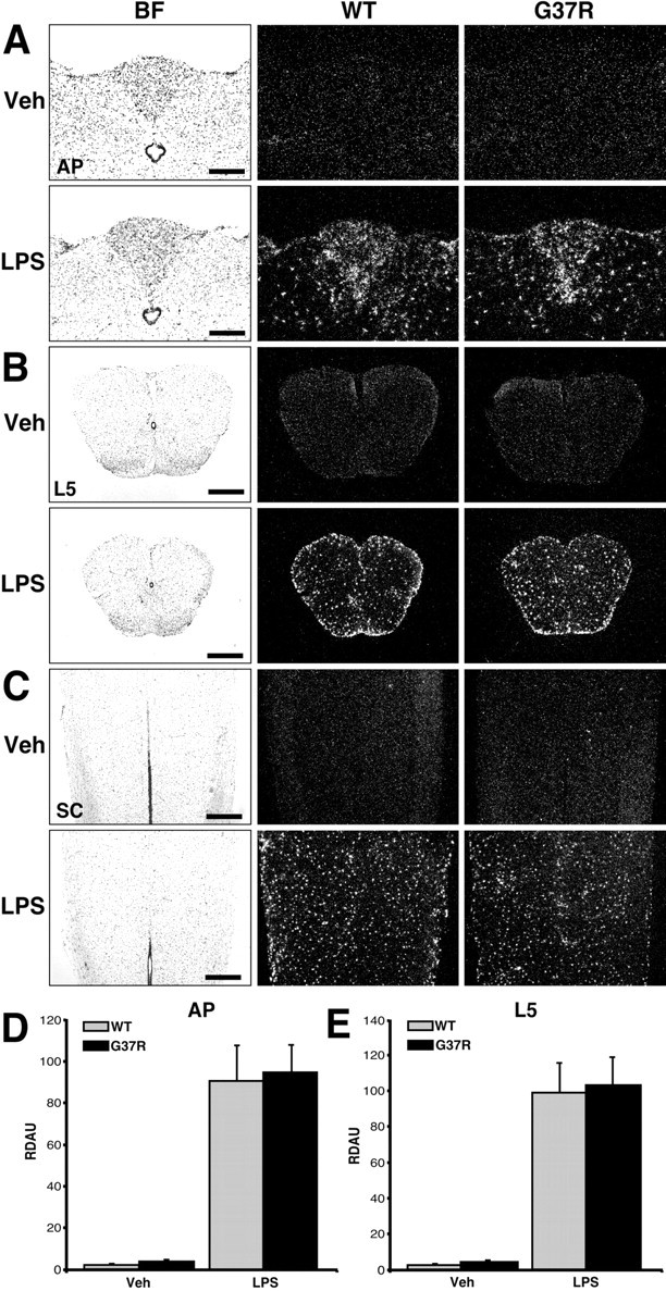
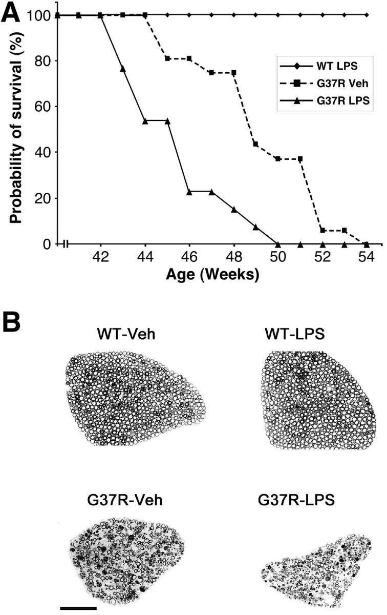
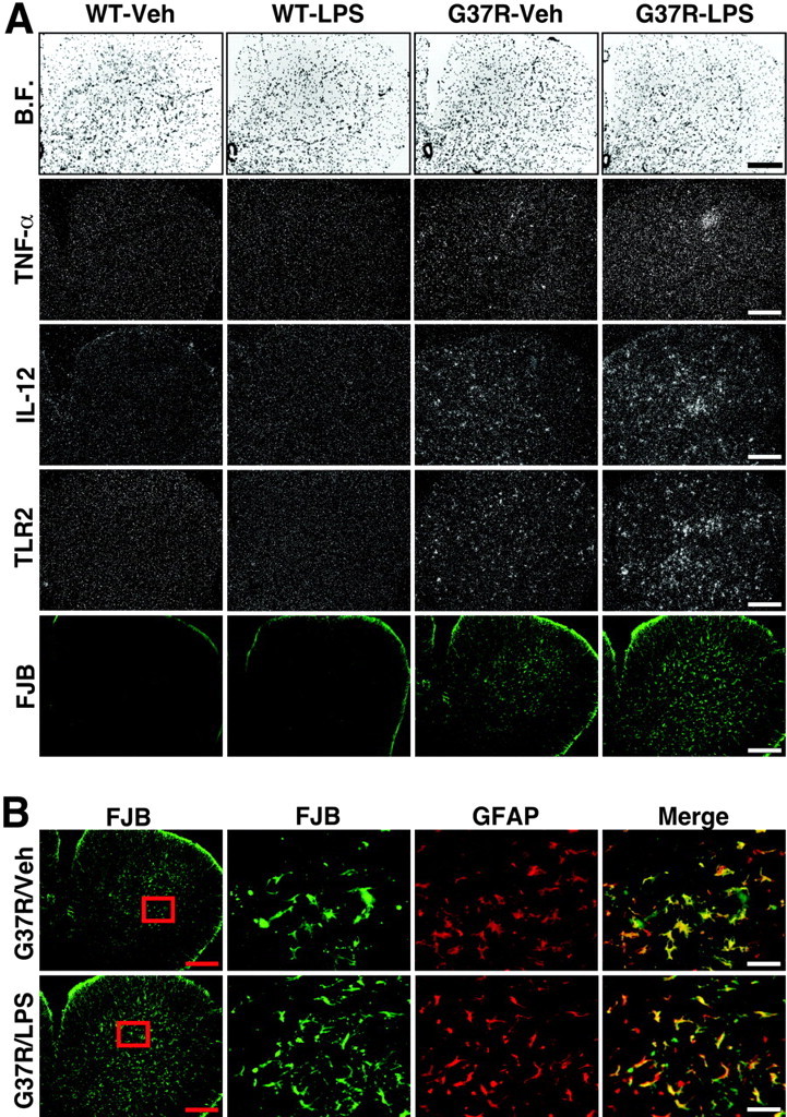
Innate immunity is a specific and organized immunological program engaged by peripheral organs and the CNS to maintain homeostasis after stress and injury. In neurodegenerative disorders, its putative deregulation, featured by inflammation and activation of glial cells resulting from inherited mutations or viral/bacterial infections, likely contributes to neuronal death. However, it remains unclear to what extent environmental factors and innate immunity cooperate to modulate the interactions between the neuronal and non-neuronal elements in the perturbed CNS. In the present study, we addressed the effects of acute and chronic administration of lipopolysaccharide (LPS), a Gram-negative bacterial wall component, in a genetic model of neurodegeneration. Transgenic mice expressing a mutant form of the superoxide dismutase 1 (SOD1(G37R)) linked to familial amyotrophic lateral sclerosis were challenged intraperitoneally with a single nontoxic or repeated injections of LPS (1 mg/kg). At different ages, SOD1(G37R) mice responded normally to acute endotoxemia. Remarkably, only a chronic challenge with LPS in presymptomatic 6-month-old SOD1(G37R) mice exacerbated disease progression by 3 weeks and motor axon degeneration. Closely associated with the severity of disease is the stronger and restricted upregulation of the receptor of innate immunity Toll-like receptor 2 and proinflammatory cytokines in degenerating regions of the ventral spinal cord and efferent fiber tracts of the brain from the LPS-treated SOD1(G37R) mice. This robust immune response was not accompanied by the establishment of acquired immunity. Our results provide solid evidence that environmental factors and innate immunity can cooperate to influence the course of disease of an inherited neuropathology.


Figures

Figure 1.
Figure 1.
Effect of a single bolus of LPS on TLR2 gene expression in the brains and spinal cords of SOD1G37R mice and their WT littermates. These bright-field (BF) and dark-field photomicrographs depict the expression pattern of TLR2 mRNA 24 hr after a single intraperitoneal injection of vehicle (Veh) or LPS (1 mg/kg of body weight). Coronal and longitudinal sections were hybridized using a mouse TLR2 cRNA probe and dipped into NTB2 emulsion milk. A, Coronal sections at the level of the AP. B, Coronal sections within the L5 segment of the spinal cord (SC). C, Longitudinal slices of the SC. Note the strong and similar hybridization signal within the brain and spinal cord of both WT and SOD1G37R mice that received an intraperitoneal bolus of LPS. Semiquantitative analysis was performed in regions of the AP (D) and L5 segment (E). Data are means ± SEM. The expression levels were comparable in the CNS of both mouse strains after the acute endotoxemia. Statistical analysis was performed by a two-way ANOVA, which indicated a significant main effect (p < 0.0001) between the vehicle- and LPS-treated groups. Scale bars: A, 200 μm; B, C, 500 μm. RDAU, Refraction density in arbitrary units. (Means ± SEM).
Figure 3.
Figure 3.
Chronic treatment with the endotoxin LPS increases the innate immune response and neurodegeneration in SOD1G37R mice. The bright-field (B.F.) and dark-field photomicrographs depict representative examples of the hybridization signal for TNF-α (B), IL-12 (C), and TLR2 (D) mRNA in the reticular formation just above the olivary complex. It is of interest to note that the hybridization signal for IL-12 and TLR2 overlaps with the fluorochrome FJB (C, D;vs E), used here as a marker of neuronal death. Degenerating axons were labeled by FJB staining in the reticular formation; DAPI-positive nuclei were essentially devoid of FJB signal in this structure (F). The bottom panels in G depict examples of microglial cells containing positive hybridization signal for TLR2 mRNA in the reticular formation. Cells of myeloid origin were labeled by immunoperoxidase using antisera directed against iba1 (brown immunoreactive cells). TLR2 mRNA was thereafter hybridized on the same sections by means of a radioactive in situ hybridization technique (silver grains). Note the presence of the mRNA encoding TLR2 within parenchymal microglia (agglomeration of silver grains within the cell cytoplasm) (black arrowheads). Scale bars: A-E, 500 μm; F, G, 50 μm. See the legend to Figure 2 for definitions of abbreviations.
Figure 2.
Figure 2.
Exacerbation of motor axon degeneration in chronically LPS-treated SOD1G37R mice accelerates disease progression. A, Survival curves of transgenic mice expressing SOD1G37R challenged systemically with LPS or vehicle every 2 weeks. Disease progression of chronically LPS-treated mice is exacerbated by ∼3 weeks. Note that the life span of wild-type mice is unaffected by the same dose of LPS (see Materials and Methods). The survival probability of transgenic mice is plotted as a function of their age in weeks. B, Transverse sections of L5 ventral root from normal mice treated chronically with vehicle (WT-Veh) or LPS (WT-LPS) and from SOD1G37R mice challenged chronically with vehicle (G37R-Veh) or LPS (G37R-LPS). Massive degeneration is observed in the L5 ventral root of G37R-Veh. A more severe loss of motor axons is found in the L5 ventral root of G37R-LPS. It is noteworthy that WT-LPS mice do not show any sign of neurodegeneration. Scale bar, 100 μm.
Figure 4.
Figure 4.
Robust inflammatory response in ventral spinal horn of chronically LPS-treated SOD1G37R mice associated with massive degeneration of astrocytes. The bright-field (B.F.) and dark-field photomicrographs depict representative examples of the hybridization signal for TNF-α, IL-12, and TLR2 mRNA in the L5 segment of the spinal cord (A). Here also the hybridization signal for IL-12 and TLR2 overlaps with the fluorochrome FJB. Neuronal cell bodies and axons contained FJB staining in this region. Please also note the robust FJB signal over astrocytes, a phenomenon that was specific to the L5 region (B). These data suggest an intimate link between degeneration of neurons and astrocytes in this region of spinal cord from SOD1G37R mice. Age of mice at time of analysis, 45-46 weeks. Scale bars: A and B (left panels), 500μm; B (high magnifications, merge), 50μm. See the legend to Figure 2 for definitions of abbreviations.
Figure 5.
Figure 5.
Relative expression levels of TLR2 hybridization signal in the brains of SOD1G37R mice and their wild-type littermates that received chronic systemic injections of the endotoxin LPS or sterile saline solution (Veh). The selected structures were the areas adjacent to the mesencephalic nucleus of the trigeminal (A), the facial nucleus (B), and the reticular formation just above the olivary complex (C). These regions were chosen to facilitate the analysis among animals, although the hybridization signal was not limited to these specific nuclei (see Results). The signals revealed on dipped NTB2 nuclear emulsion slides were analyzed and quantified (relative levels) with an Olympus Optical System (BX-50; BMax) coupled to a Macintosh computer (PowerPC 7100/66) and Image software [version 1.59; non-FPU; W. Rasband (National Institutes of Health, Bethesda, MD)]. The refraction density in arbitrary units (RDAU) of the hybridization signal was measured under dark-field illumination at a magnification of 10×. Sections from experimental and control animals were digitized and subjected to densitometric analysis, yielding measurements of RDAU. The RDAU of each region was then corrected for the average background signal, which was determined by sampling cells immediately outside the cell group of interest. Data are reported as mean values (±SEM) for vehicle- and LPS-treated animals of both mouse stains. Statistical analysis was performed by a two-way ANOVA, followed by a Bonferroni-Dunn test procedure as post hoc comparisons by means of the Statview program (version 4.01; Macintosh). *Significantly different (p < 0.05) from WT groups of mice. **Significantly different (p < 0.05) from all of the other groups. Age of mice at time of analysis, 45-46 weeks.
Figure 6.
Figure 6.
Acute and chronic administration of LPS in SOD1G37R failed to alter expression of endogenous and transgene SOD1 Six-month-old WT and transgenic SOD1G37R littermates were analyzed for SOD1 levels 24 hr after acute injection of LPS (1 mg/kg of body weight). Expression of both endogenous mSOD1 and hSOD1 remained unaffected in spinal cord (A, lanes 5-8) and spleen (B, lanes 5-8) of SOD1G37R animals in response to saline (Veh) or LPS injection as detected by means of an antibody recognizing both SOD1 proteins. The endotoxin also failed to significantly upregulate mSOD1 expression in WT animals (A, B, lanes 1-4). Similar levels of both SOD1s were found in WT and SOD1G37R mice 48 hr after LPS or Veh administration (data not shown). In SOD1G37R mice that were chronically treated with LPS, expression of mutant SOD1 (detected with an antibody directed against the human transgene) remained stable when compared with littermates treated with Veh (C). Lysates from SOD1G37R line 42 (L42) overexpressing 2- to 2.5-fold the levels of line 29 (L29) were used as comparative control for expression levels. Thus, neither acute nor chronic administration of LPS in SOD1G37R mice affected expression of endogenous and transgene SOD1. Actin and α-tubulin were used as controls for loadings. Quantifications were corrected with levels of actin or tubulin, and performed with the Labscan program. Two to 4 animals were used for each condition. Experiments were repeated from three to eight times. Results represent means ± SD for all of the experiments and animals. See the legend to Figure 2 for definitions of abbreviations.

References

    1. Akira S, Takeda K, Kaisho T (2001) Toll-like receptors: critical proteins linking innate and acquired immunity. Nat Immunol 2: 675-680. - PubMed
    1. Aliprantis AO, Yang RB, Mark MR, Suggett S, Devaux B, Radolf JD, Klimpel GR, Godowski P, Zychlinsky A (1999) Cell activation and apoptosis by bacterial lipoproteins through Toll-like receptor-2. Science 285: 736-739. - PubMed
    1. Aliprantis AO, Yang RB, Weiss DS, Godowski P, Zychlinsky A (2000) The apoptotic signaling pathway activated by Toll-like receptor-2. EMBO J 19: 3325-3336. - PMC - PubMed
    1. Bieber AJ, Warrington A, Pease LR, Rodriguez M (2001) Humoral autoimmunity as a mediator of CNS repair. Trends Neurosci 24: S39-S44. - PubMed
    1. Bristol LA, Rothstein JD (1996) Glutamate transporter gene expression in amyotrophic lateral sclerosis motor cortex. Ann Neurol 39: 676-679. - PubMed

Publication types

MeSH terms