. 2004 Feb 11;24(6):1393-7.
doi: 10.1523/JNEUROSCI.4986-03.2004.

A novel DNA enzyme reduces glycosaminoglycan chains in the glial scar and allows microtransplanted dorsal root ganglia axons to regenerate beyond lesions in the spinal cord

Affiliations

A novel DNA enzyme reduces glycosaminoglycan chains in the glial scar and allows microtransplanted dorsal root ganglia axons to regenerate beyond lesions in the spinal cord

Barbara Grimpe et al. J Neurosci. .

Abstract

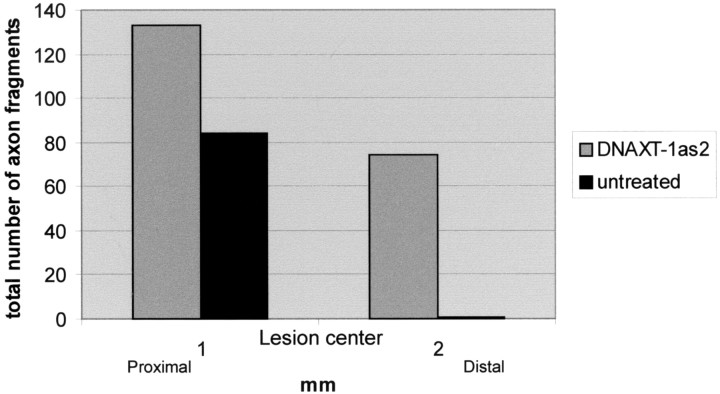
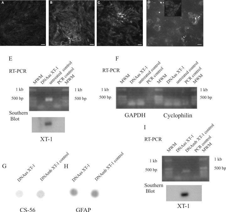
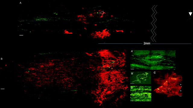
CNS lesions induce production of ECM molecules that inhibit axon regeneration. One major inhibitory family is the chondroitin sulfate proteoglycans (CSPGs). Reduction of their glycosaminoglycan (GAG) chains with chondroitinase ABC leads to increased axon regeneration that does not extend well past the lesion. Chondroitinase ABC, however, is unable to completely digest the GAG chains from the protein core, leaving an inhibitory "stub" carbohydrate behind. We used a newly designed DNA enzyme, which targets the mRNA of a critical enzyme that initiates glycosylation of the protein backbone of PGs, xylosyltransferase-1. DNA enzyme administration to TGF-beta-stimulated astrocytes in culture reduced specific GAG chains. The same DNA enzyme applied to the injured spinal cord led to a strong reduction of the GAG chains in the lesion penumbra and allowed axons to regenerate around the core of the lesion. Our experiments demonstrate the critical role of PGs, and particularly those in the penumbra, in causing regeneration failure in the adult spinal cord.


Figures

Figure 2.
Figure 2.
The histogram shows the numbers of microtransplanted DRG axon fragments proximal and distal to the lesion in DNA enzyme-treated and untreated spinal cords.
Figure 1.
Figure 1.
Confocal photomicrographs of TGF-β-stimulated primary astrocyte cultures treated with the DNA enzyme (A) or control DNA enzyme (B) or left untreated (C) and stained with the CS-56 antibody. Uptake of the biotin-streptavidin-labeled DNA enzyme can be seen in the astrocytes (D). Scale bars, 10 μm. E, F, RT-PCR-Southern blot of primary astrocyte cultures for XT-1 (E) and RT-PCR for GAPDH and cyclophilin (F) of the same mRNA. G, Dot blot of primary astrocyte cultures treated with the DNA enzyme or control DNA enzyme and stained with the CS-56 antibody. H, Restaining of the same dot blot with GFAP. I, RT-PCR-Southern blot for the XT-1 in a DNA enzyme- and control DNA enzyme-treated spinal cord.
Figure 3.
Figure 3.
Confocal photomicrographs of treated and untreated spinal cords. Red/green (A) and green (A′) channels show the green DRGs and their axons passing the distinct borders of the lesion cores (red CS-56 staining) in a DNA enzyme-treated spinal cord. Axons that enter the lesion core become dystrophic (A, A′, arrows). Red/green (B) and green (B′) channels show an untreated spinal cord with the green DRG and red CS-56 staining. Note the struggling axon in the heart of the lesion. Scale bar, 50 μm. C, Diffusion of a labeled DNA enzyme in vivo 7 d after treatment. Scale bar, 100 μm. D, GFAP-stained section of the lesion area in a different animal. Note the disrupted astrocyte alignment in the core and borders of the lesion (L) and the aligned astrocytes (arrows) in the region where axons regenerate.

Similar articles

Cited by

References

    1. Altschult SF, Madden TL, Schaeffer AA, Zhang J, Zhang Z, Miller W, Lipman DJ (1997) Gapped BLAST and PSI-BLAST: a new generation of protein database search programs. Nucleic Acids Res 25: 3389-3402. - PMC - PubMed
    1. Asher RA, Morgenstern DA, Fidler PS, Adcock KH, Oohira A, Braistead JE, Levine JM, Margolis RU, Rogers JH, Fawcett JW (2000) Neurocan is upregulated in injured brain and in cytokine-treated astrocytes. J Neurosci 20: 2427-2438. - PMC - PubMed
    1. Bradbury EJ, Moon LD, Popat RJ, King VR, Bennett GS, Patel PN, Fawcett JW, McMahon SB (2002) Chondroitinase ABC promotes functional recovery after spinal cord injury. Nature 416: 636-640. - PubMed
    1. Brose K, Bland KS, Wang KH, Arnott D, Henzel W, Goodman CS, Tessier-Lavigne M, Kidd T (1999) Slit proteins bind Robo receptors and have an evolutionarily conserved role in repulsive axon guidance. Cell 96: 795-806. - PubMed
    1. Bundesen LQ, Scheel TA, Bregman BS, Kromer LF (2003) Ephrin-B2 and EphB2 regulation of astrocyte-meningeal fibroblast interactions in response to spinal cord lesions in adult rats. J Neurosci 23: 7789-7800. - PMC - PubMed

Publication types

MeSH terms