Comparative Study
. 2004 Feb;34(2):303-19.
doi: 10.1016/j.bone.2003.10.005.

MEPE has the properties of an osteoblastic phosphatonin and minhibin

Affiliations
Comparative Study

MEPE has the properties of an osteoblastic phosphatonin and minhibin

P S N Rowe et al. Bone. 2004 Feb.

Abstract

Matrix extracellular phosphoglycoprotein (MEPE) is expressed exclusively in osteoblasts, osteocytes and odontoblasts with markedly elevated expression found in X-linked hypophosphatemic rickets (Hyp) osteoblasts and in oncogenic hypophosphatemic osteomalacia (OHO) tumors. Because these syndromes are associated with abnormalities in mineralization and renal phosphate excretion, we examined the effects of insect-expressed full-length human-MEPE (Hu-MEPE) on serum and urinary phosphate in vivo, (33)PO(4) uptake in renal proximal tubule cultures and mineralization of osteoblast cultures. Dose-dependent hypophosphatemia and hyperphosphaturia occurred in mice following intraperitoneal (IP) administration of Hu-MEPE (up to 400 microg kg(-1) 31 h(-1)), similar to mice given the phosphaturic hormone PTH (80 microg kg(-1) 31 h(-1)). Also the fractional excretion of phosphate (FEP) was stimulated by MEPE [65.0% (P < 0.001)] and PTH groups [53.3% (P < 0.001)] relative to the vehicle group [28.7% (SEM 3.97)]. In addition, Hu-MEPE significantly inhibited (33)PO(4) uptake in primary human proximal tubule renal cells (RPTEC) and a human renal cell line (Hu-CL8) in vitro (V(max) 53.4% inhibition; K(m) 27.4 ng/ml, and V(max) 9.1% inhibition; K(m) 23.8 ng/ml, respectively). Moreover, Hu-MEPE dose dependently (50-800 ng/ml) inhibited BMP2-mediated mineralization of a murine osteoblast cell line (2T3) in vitro. Inhibition of mineralization was localized to a small (2 kDa) cathepsin B released carboxy-terminal MEPE peptide (protease-resistant) containing the acidic serine-aspartate-rich motif (ASARM peptide). We conclude that MEPE promotes renal phosphate excretion and modulates mineralization.


Figures

Fig. 1
Fig. 1
Key features and localization of the acidic serine–aspartate-rich MEPE-associated motif (ASARM peptide and motif). (A) Scheme showing the last 50-amino acid COOH-terminal residues of MEPE in man, mouse and rat, respectively. The ASARM motif is highly conserved in man, macaque monkey, mouse and rat and is localized to the last COOH-terminal 18-amino acid residues of the large, approximately 500-residue MEPE proteins as depicted in the scheme [71]. Cathepsin B (an osteoblast protease) specifically cleaves MEPE at the COOH terminus releasing ASARM peptide. The cathepsin B cleavage site does not occur elsewhere in MEPE and is highly conserved between species. (B) Moreover, the ASARM peptide is uniquely resistant to many proteases (trypsin, papain, proteinase K, carboxypeptidases, tryptase, etc.). The ASARM motif is found in members of the SIBLING protein family (MEPE, DMP-1, osteopontin, DSPP) and in osteopontin occurs in the mid-region of the molecule [71] and also salivary statherin. The dark boxes represent the position of the ASARM motif in MEPE and osteopontin and the number of amino acid residues for each respective protein is indicated [71]. Recently, Hoyer et al. [32] demonstrated that the osteopontin ASARM motif [71] inhibits calcium oxalate crystallization and growth. Also, in vivo studies using hyperoxaluria induced osteopontin knock-out mice have confirmed that osteopontin/osteopontin peptides are critical inhibitors of renal stone formation [96] and mineral maturity (mineral crystal size and perfection) throughout all anatomic regions of the osteopontin knock-out mouse bone is significantly increased [10].
Fig. 2
Fig. 2
Scheme illustrating a proposed role and molecular mechanism for PHEX, MEPE and the ASARM peptide in mineralization. Cathepsin B is expressed in the osteoblast together with PHEX and MEPE [1,2,16,26,28,29,50,61,71,75,86]. NEP, ECEL-1/DINE and cathepsin D and MEPE are up-regulated in Hyp osteoblasts that have defective PHEX [2,3,17,29,34]. PHEX protects MEPE from cathepsin B specific cleavage [29] (possibly by sequestration on the cell surface) and prevents release of the ASARM peptide. Thus, in rickets, increased levels of ASARM peptide are proposed to inhibit mineralization (defective PHEX). Also, the ASARM peptide is resistant to a vast range of proteases due to its unusual sequence. The MEPE ASARM peptide (NH2-FSSRRRDDSSESSDSGSSSESDGD-COOH) [71] inhibits mineralization in vivo (this manuscript) and the osteopontin ASARM peptide (NH2-DDSHQSDESHHSDESDED-COOH) [32,71] potently inhibits calcium oxalate crystallization and crystal growth [32]. Also, the salivary statherin ASARM peptide contains the specific peptide sequence shown to play a biological role in inhibiting spontaneous precipitation of supersaturated salivary calcium and phosphate and maintaining the mineralization dynamics of tooth enamel [5,43,64,78]. The MEPE knock-out (complete absence of MEPE ASARM peptide and MEPE) as expected has accelerated mineralization, increased bone density and bone formation [26]. Moreover, studies in vivo using hypoxularia renal stone induction in osteopontin null-mutant mice confirm that osteopontin/osteopontin peptides are critical inhibitors of renal stone formation [96]. Mineral maturity (mineral crystal size and perfection) throughout all anatomic regions of the osteopontin knock-out mouse bone is also significantly increased [10]. Of additional interest is the recent finding that loss of function mutations in the cathepsin C gene is the primary cause of Papillon–Lefevre syndrome (PLS) [88]. PLS results in periodontopathia with loss of both deciduous and permanent dentitions and severe intracranial calcifications [88]. MEPE is abundantly expressed in both brain (man/primates) and odontoblasts/osteoblasts (rodents and man/primates) [26,45,71]. Confirmation of this model requires further studies.
Fig. 3
Fig. 3
SDS-polyacrylamide electrophoretic separation and Western blotting of purified insect-expressed full-length human-MEPE (Hu-MEPE). A shows a Coomassie-stained gel with three lanes containing bovine serum albumin (BSA), a single lane of standard protein molecular weight markers (Ma) and two lanes containing MEPE. B shows the corresponding PVDF membrane blot containing immobilized proteins screened with mid-region (RGD peptide) MEPE polyclonal (see Materials and methods section). Confirmation of MEPE identity was achieved by N-terminal amino acid sequence analysis of the excised PVDF blot region corresponding to the positive Western band. Purified protein contained N-terminal amino acid residues APTFQ confirming cleavage of predicted nascent-MEPE signal peptide by the sf9 insect cells.
Fig. 4
Fig. 4
Dose-dependent in vitro inhibition of 33PO4 uptake is induced by Hu-MEPE in primary human proximal tubule epithelial cells (RPTEC) and a human renal cell line (Hu-CL8). A and B graphically illustrate the inhibition using Lineweaver–Burk plots [inverse percentage rate of inhibition against the inverse MEPE concentration (ng/ml)]. The Hu-CL8 cell line (A) and human RPTEC primary renal cells (B) have similar Km values (23.8 and 27.39 ng/ml, respectively) but differing Vmax values (9.1% and 53.4%, respectively). Graph C (histogram) illustrates Hu-MEPE and PTH dose-dependent inhibition observed with RPTEC cells in vitro (see text for discussion). Normal; buffer solvent, MEPE-25 (25 ng/ml), MEPE-50 (50 ng/ml), MEPE-100 (100 ng/ml), PTH-10 (10 ng/ml), PTH-100 (100 ng/ml). Differences in C were assessed statistically by the use of Newman–Keuls multiple comparison equations after ANOVA (non-parametric). A P value of less than 0.05 was considered significant. The SEM was used as a representative measure of how far the sample mean differed from the true population mean (see bars). For A, B and C, each data point contained N = 12 and N = 15 replicates for MEPE and PTH, respectively.
Fig. 5
Fig. 5
MEPE and PTH induce hypophosphatemia and increase the fractional excretion of phosphate (FEP) when administered intraperitoneally (IP). (A) Histogram of serum phosphate measured after 31 h and four bolus IP injections of MEPE40 (40 μg kg–1 30 h–1), MEPE400 (400 μg kg–1 30 h–1) and PTH (80 μg kg–1 30 h–1). (B and C) Histograms illustrating the changes in FEP after 7 h (three bolus injections IP) and 31 h (four bolus injections IP). See Materials and methods for calculations and detailed description of protocol. Differences were assessed statistically by the use of Newman–Keuls multiple comparison equations after ANOVA (non-parametric). A P value of less than 0.05 was considered significant. The SEM was used as a representative measure of how far the sample mean differed from the true population mean (see bars) and each group contained N = 7 animals.
Fig. 6
Fig. 6
Hu-MEPE and MEPE ASARM peptide (CFSSRRRDDSSESSDSGSSSESDGD) inhibit BMP2-mediated mineralization of mouse osteoblast cell line 2T3. Wells were stained for mineralization nodule formation using von Kossa (see Materials and methods) and the results after 26 days culture are shown. Row A: upper two wells (BMP2), contain BMP2 (100 ng/ml); middle two wells (control), control cells with no BMP2 or peptide; lower two wells (BMP2 and control peptide), BMP2 (100 ng/ml) with control peptide (CGSGYTDLQERGDNDISPFSGDGQPF) at 300 ng/ml (108.6 pmol/ml). Row B: Upper two wells (BMP2 and MEPE 100), contain Hu-MEPE (100 ng/ml) plus BMP2 (100 ng/ml); middle two wells (BMP2 and MEPE 500), Hu-MEPE (500 ng/ml) plus human-BMP2 (100 ng/ml); lower two wells (BMP2 and MEPE 800), MEPE (800 ng/ml) plus BMP2 (100 ng/ml). Cells in which MEPE was added in the absence of BMP2 (data not shown) were indistinguishable to control cells (Row A, middle two wells). Row C: Upper two wells (BMP2 and ASARM 60), BMP2 (100 ng/ml) plus MEPE ASARM peptide (CFSSRRRDDSSESSDSGSSSESDGD) at 60 ng/ml (22.7 pmol/ml); lower two wells (BMP2 and ASARM 300), BMP2 (100 ng/ml) plus MEPE ASARM peptide at 300 ng/ml (113.5 pmol/ml).
Fig. 7
Fig. 7
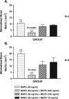
Quantification of mineralization inhibition of mouse osteoblast 2T3 cell line by Hu-MEPE as assessed by von Kossa staining. Concentrations above 100 ng/ml completely inhibited mineralization (see Fig. 6). Thus, effects at 10, 100 and 500 ng/ml MEPE are shown after day 13 (A) and day 20 (B), respectively. The mineralized bone matrix formation of 2T3 cells were quantitated by computer image analysis as previously described [14] and the data represent the mean for three samples (see Materials and methods). ANOVA non-parametric and Neuman–Keuls multiple comparison confirm highly significant inhibition of BMP2 mediated mineralization by Hu-MEPE (see graph). The SEM was used as a representative measure of how far the sample mean differed from the true population mean (see bars).
Fig. 8
Fig. 8
Salivary statherin and MEPE consensus ASARM motif: mineralization inhibition and ancestral genes on chromosome 4. The depicted scheme illustrates the remarkable association of MEPE, DMP-1 and the SIBLINGs to an ancestral mineralization gene that is also thought to play a key role in phosphate calcium transport in saliva (salivary statherin). Statherin maps to chromosome 4 in the SIBLING/MEPE region and also contains an ASARM motif. Statherin is a 62-residue peptide with asymmetric charge and structural properties [43,64]. The upper scheme (A) depicts a clustal alignment of the COOH-terminal region of human-DMP-1 human-MEPE, mouse-MEPE and rat-MEPE with human-Statherin. In MEPE, the ASARM peptide is the most distal region of the molecule encompassing the last 17 residues of the COOH terminus and the region is highlighted with a boxed cartouche labeled MEPE ASARM peptide (A). In DMP-1, the ASARM region is also at the carboxy terminus but ends at residue 480 slightly upstream of the distal COOH terminus (protein 513 residues long). The short 62-residue statherin molecule contains an ASARM motif region as depicted in the diagram and the key residues are highlighted in the consensus string shown at the bottom of A. The boxed cartouche labeled as statherin ASARM peptide contains the specific statherin ASARM peptide sequence shown to play a biological role in inhibiting spontaneous precipitation of supersaturated salivary calcium and phosphate and maintaining the mineralization dynamics of tooth enamel [5,43,64,78]. As with the MEPE ASARM peptide, a single cathepsin B site is present in statherin that would potentially release the highly charged and phosphorylated aspartate–serine-rich statherin ASARM peptide [indicated by line between statherin arginine (R) residues 29 and 30; (A). In Statherin, the cathepsin B cleavage site is adjacent and located COOH-terminal to the motif. In MEPE, the cathepsin B cleavage site is also adjacent to the ASARM motif but asymmetrically arranged NH2-terminal to the motif between the arginine and aspartate. In both cases (MEPE and statherin), cleavage would result in the release of a short phosphorylated aspartate/serine-rich acidic peptide of low pI and almost identical physiocochemical properties. A feature of the MEPE ASARM region is the repeat (D) SSES/E sequence. This short sequence has been shown to be key inhibitor of hydroxapatite crystals formation and mineralization in salivary statherin [43,64]. The MEPE ASARM region is highly homologous to the DMP-1 but the single cathepsin B site in DMP-1 is located further upstream toward the NH2 terminus (A). B schematically presents the remarkable clustering of MEPE, DMP-1, statherin and other SIBLING genes on chromosome 4. All contain an ASARM motif in differing structural contexts with many diverse structural and genomic features (exon–intron structure) and associations with bone dental functions.

References

    1. Aisa MC, Rahman S, Senin U, Maggio D, Russell RG. Cathepsin B activity in normal human osteoblast-like cells and human osteoblastic osteosarcoma cells (MG-63): regulation by interleukin-1 beta and parathyroid hormone. Biochim Biophys Acta. 1996;1290:29–36. - PubMed
    1. Argiro L, Desbarats M, Glorieux FH, Ecarot B. Mepe, the gene encoding a tumor-secreted protein in oncogenic hypophosphatemic osteomalacia, is expressed in bone. Genomics. 2001;74:342–51. - PubMed
    1. Bai X, Miao D, Panda D, Grady S, McKee MD, Goltzman D, et al. Partial rescue of the Hyp phenotype by osteoblast-targeted PHEX (phosphate-regulating gene with homologies to endopeptidases on the X chromosome) expression. Mol Endocrinol. 2002;16:2913–25. - PubMed
    1. Bai XY, Miao D, Goltzman D, Karaplis AC. The autosomal dominant hypophosphatemic rickets R176Q mutation in FGF23 resists proteolytic cleavage and enhances in vivo biological potency. J Biol Chem. 2003;7:7. - PubMed
    1. Bennick A, McLaughlin AC, Grey AA, Madapallimattam G. The location and nature of calcium-binding sites in salivary acidic pro-line-rich phosphoproteins. J Biol Chem. 1981;256:4741–6. - PubMed

Publication types

MeSH terms

LinkOut - more resources