. 2004 Mar;78(5):2277-87.
doi: 10.1128/jvi.78.5.2277-2287.2004.

Role for CCR5Delta32 protein in resistance to R5, R5X4, and X4 human immunodeficiency virus type 1 in primary CD4+ cells

Affiliations

Role for CCR5Delta32 protein in resistance to R5, R5X4, and X4 human immunodeficiency virus type 1 in primary CD4+ cells

Lokesh Agrawal et al. J Virol. 2004 Mar.

Abstract

CCR5Delta32 is a loss-of-function mutation that abolishes cell surface expression of the human immunodeficiency virus (HIV) coreceptor CCR5 and provides genetic resistance to HIV infection and disease progression. Since CXCR4 and other HIV coreceptors also exist, we hypothesized that CCR5Delta32-mediated resistance may be due not only to the loss of CCR5 function but also to a gain-of-function mechanism, specifically the active inhibition of alternative coreceptors by the mutant CCR5Delta32 protein. Here we demonstrate that efficient expression of the CCR5Delta32 protein in primary CD4(+) cells by use of a recombinant adenovirus (Ad5/Delta32) was able to down-regulate surface expression of both wild-type CCR5 and CXCR4 and to confer broad resistance to R5, R5X4, and X4 HIV type 1 (HIV-1). This may be important clinically, since we found that CD4(+) cells purified from peripheral blood mononuclear cells of individuals who were homozygous for CCR5Delta32, which expressed the mutant protein endogenously, consistently expressed lower levels of CXCR4 and showed less susceptibility to X4 HIV-1 isolates than cells from individuals lacking the mutation. Moreover, CD4(+) cells from individuals who were homozygous for CCR5Delta32 expressed the mutant protein in five of five HIV-exposed, uninfected donors tested but not in either of two HIV-infected donors tested. The mechanism of inhibition may involve direct scavenging, since we were able to observe a direct interaction of CCR5 and CXCR4 with CCR5Delta32, both by genetic criteria using the yeast two-hybrid system and by biochemical criteria using the coimmunoprecipitation of heterodimers. Thus, these results suggest that at least two distinct mechanisms may account for genetic resistance to HIV conferred by CCR5Delta32: the loss of wild-type CCR5 surface expression and the generation of CCR5Delta32 protein, which functions as a scavenger of both CCR5 and CXCR4.


Figures
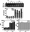
FIG. 1.
FIG. 1.
CD4+ cells from individuals homozygous for CCR5Δ32 (−/−) are resistant to infection with both X4 and R5 HIV. (A) The genotypes of the PBMC samples were verified by RT-PCR amplification of a fragment that spans the 32-bp deletion. CD4+ cells purified from four −/− and four +/+ individuals were stimulated with PHA plus IL-2 for 3 days and were used in an HIV-1 infection assay (B) or for FACS analysis of CXCR4 (C) or CD4 (D). The p24 values in panel B represent amounts produced at day 9 postinfection. The results shown are from a single experiment representative of four independent experiments. MFI, mean fluorescence intensity; M, DNA size marker.
FIG. 2.
FIG. 2.
CCR5+ CXCR4+ double-positive cells in unstimulated and stimulated PBMCs. Three-color flow cytometry of freshly isolated PBMCs was performed on either unstimulated cells or cells stimulated with PHA plus IL-2 or αCD3 antibody plus IL-2. PBMCs were gated according to forward and side scatter and with either CD3-cychrome (HIT3Aa) or CD4-cychrome (RPA-T4) staining. The two-dimensional plots show expression of CCR5 (fluorescein isothiocyanate conjugate; 2D7 MAb) versus CXCR4 (PE conjugate; 12G5 MAb). Percentages of cells that are positive in the respective quadrants are indicated. Results for the respective isotype control antibodies are shown in the top two plots for each donor. The cell treatment is indicated to the right of each pair of plots, and gating is indicated at the bottom of each column of plots. The figure shows primary data from two different individuals and Table 1 summarizes the results from these two donors and eight others.
FIG. 3.
FIG. 3.
Detection of endogenous CCR5Δ32 protein in unstimulated primary CD4+ cells from CCR5Δ32 homozygotes. (A) Genotypes were confirmed by RT-PCR amplification of the appropriate fragment of CCR5 (648 bp) or CCR5Δ32 (616 bp) from purified CD4+ cells from −/− and +/+ individuals. −/−, the genotype of individuals homozygous for the CCR5Δ32 allele; +/+, individuals homozygous for the wt CCR5 allele. The CD4+ cells were purified from five HIV-uninfected (−/−) donors (identified as UN 1, 2, and 3 in panel B and 4 and 5 in panel C), two different HIV-infected (−/−) individuals (labeled IN 1 and 2), and three HIV-uninfected individuals lacking CCR5Δ32. The CCR5Δ32 protein was detected by using anti-CCR5Δ32 (anti-Δ32), a polyclonal rabbit antiserum directed against the novel frame-shifted amino acids that specifically recognizes the mutant protein (B), or antibodies against the common N terminus of CCR5 and CCR5Δ32 proteins (C). Probing similar blots with preimmune serum did not show any protein bands specific for the CCR5Δ32 protein (not shown). (D) Up-regulation of CXCR4 and down-regulation of CCR5Δ32 proteins upon PHA plus IL-2 stimulation of CD4+ cells (Stim). Unstim., unstimulated. The numbers at the left in panel C indicate the positions of protein standards. kDa, kilodaltons; M, DNA size marker.
FIG. 4.
FIG. 4.
Expression of CCR5Δ32 protein in 293 cells. (A) Structural map of a recombinant adenovirus (Ad5) encoding the Δ32 ORF. The CCR5Δ32 ORF, cloned from an HIV-seronegative CCR5Δ32 homozygote (−/−), is under the control of the major late promoter of adenovirus type 2 and the simian virus 40 poly(A) 3′ (SV40 PA) processing signal. The adenovirus genome is represented as 100 map units (m.u.), with 365 bp/m.u. (B and C) Detection of Ad5-encoded CCR5Δ32 protein in infected 293 cells. Ad5-encoded CCR5Δ32 protein was detected by using anti-CCR5Δ32 antisera (anti-Δ32) directed against the frame-shifted domain of CCR5Δ32 (B) or antisera directed against the CCR5 N terminus (anti-CCR5) (C). Note that bands above the 34-kDa marker band are frequently detected with anti-CCR5Δ32 but not with anti-CCR5 antibodies. (D) Comparison of CCR5Δ32 protein levels made in Ad5/Δ32-infected CD4+ cells with those endogenously made in −/− CD4+ cells. The blot was reprobed with antibodies against GAPDH to control for gel loading. PBMC samples from individuals homozygous for the CCR5Δ32 allele are referred to as −/−, and those from individuals homozygous for the wt CCR5 allele are referred to as +/+. Samples labeled as 1 and 2 are the same as samples 1 and 2 used in other figures. The numbers at the left indicate the positions of protein standards. kDa, kilodaltons.
FIG. 5.
FIG. 5.
Exogenous CCR5Δ32 specifically inhibits endogenous CCR5 and CXCR4 expression and HIV coreceptor activity in primary CD4+ cells. (A) Purified CD4+ cells were infected with Ad5 vector, Ad5/Δ32, or Ad5/CCR5 and then stained for cell surface CCR5, CXCR4, CXCR2, or CD4. Staining of cells infected with Ad5 was considered 100% staining. The matched isotype staining was calculated to be <3%. (B) Purified CD4+ cells were infected with Ad5 vectors encoding the proteins indicated on the x axis, challenged with HeLa cells expressing either the control Unc, LAV (X4), or Ba-L (R5) HIV-1 Env, and examined for the extent of cell fusion by measuring β-Gal production. (C) Purified CD4+ cells were infected with WR (a vaccinia virus vector control that does not encode any foreign protein) or recombinant vaccinia viruses encoding the proteins indicated on the x axis and then were stained for cell surface CCR5, CXCR4, CCR2, or CD4. (D) Separate samples of CD4+ cells were infected with recombinant vaccinia viruses encoding the proteins indicated on the x axis, challenged with HeLa cells expressing the same HIV-1 Env proteins as in panel B, and examined for β-Gal production. (E) Some targets used in panel C were challenged with HeLa cells expressing the human T-cell leukemia virus type 1 Env, and the extent of cell fusion was measured similarly. In all of these experiments, fusion with Unc Env represents the background signal resulting from nonspecific cell fusion. The error bars represent replicates of the same experiment. The results shown represent 1 of 10 independent experiments using 10 different donors. MFI, mean fluorescence intensity.
FIG. 6.
FIG. 6.
Heterodimerization of CCR5Δ32 protein with CCR5 and CXCR4. (A) 293-CD4 cells were coinfected with either Ad5/CCR5 plus Ad5 or Ad5CCR5 plus Ad5/Δ32 and challenged with HeLa cells expressing the indicated HIV-1 Envs. The error bars represent replicates of the same experiment. The results shown represent one of three independent experiments. (B) Coimmunoprecipitation analysis. 293-CD4 cells were infected with adenovirus encoding CCR5 (CCR5) plus adenovirus encoding CCR5Δ32 (Δ32) or control adenovirus (Ad5), as indicated at the top of each pair of lanes. Coinfected cells were lysed and immunoprecipitated with anti-CCR5Δ32 antibodies (anti-Δ32). Immunoprecipitates were resuspended in native PFO loading sample buffer and subjected to PFO-10% PAGE and sequential immunoblot analysis with antibodies specific for CCR5Δ32, CCR5, and CXCR4, as indicated at the bottom of each pair of lanes. wt CCR5 was detected with CTC-6 MAbs that do not react with CCR5Δ32 protein. The numbers on the left indicate the positions of molecular weight markers. kDa, kilodaltons. The identity of each band is indicated to the right.
FIG. 7.
FIG. 7.
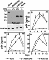
Gene delivery of CCR5Δ32 into CD4+ cells from HIV-seronegative CCR5 (+/+) individuals confers resistance to R5 and X4 HIV-1 infection. (A) Expression of CCR5Δ32 protein in CD4+ cells. Lysates of cells from individuals homozygous for wt CCR5 (+/+) or CCR5Δ32 (−/−) that had been infected with no virus (none) or adenovirus encoding CCR5 (Ad/CCR5) or CCR5Δ32 (Ad5/Δ32) were analyzed by Western blotting using anti-CCR5Δ32-specific antiserum (anti-Δ32). Note that the levels of exogenous and endogenous CCR5Δ32 protein were similar in appropriate samples. GAPDH was monitored to assess equivalent loading of samples. The numbers at the left indicate the positions of protein standards. kDa, kilodaltons. (B to D) HIV replication kinetics in CD4+ cells. Control cells were incubated with no virus (none; open squares) and compared to cells infected with either Ad5/Δ32 (open circles) or Ad5/CCR5 (open triangles) at 50 PFU/cell for each virus with regard to productive infection by the X4 HIV-1 strain IIIB (B), the R5 strain Ba-L (C), and the R5X4 strain 89.6 (D). The amount of p24 antigen in the cell-containing supernatants was measured over time by enzyme-linked immunosorbent assay. A zidovudine control infection resulted in p24 values below 1 ng/ml (not shown). The results shown are the means ± standard errors of the means from one experiment representative of five independent experiments using five different donors.

References

    1. Agrawal, L., and G. Alkhatib. 2001. Chemokine receptors: emerging opportunities for new anti-HIV therapies. Expert Opin. Ther. Targets 5:303-326. - PubMed
    1. Alkhatib, G., and D. J. Briedis. 1988. High-level eucaryotic in vivo expression of biologically active measles virus hemagglutinin by using an adenovirus type 5 helper-free vector system. J. Virol. 62:2718-2727. - PMC - PubMed
    1. Alkhatib, G., C. Combadiere, C. C. Broder, Y. Feng, P. E. Kennedy, P. M. Murphy, and E. A. Berger. 1996. CC CKR5: a RANTES, MIP-1alpha, MIP-1beta receptor as a fusion cofactor for macrophage-tropic HIV-1. Science 272:1955-1958. - PubMed
    1. Alkhatib, G., M. Locati, P. E. Kennedy, P. M. Murphy, and E. A. Berger. 1997. HIV-1 coreceptor activity of CCR5 and its inhibition by chemokines: independence from G protein signaling and importance of coreceptor downmodulation. Virology 234:340-348. - PubMed
    1. Alkhatib, G., B. Massie, and D. J. Briedis. 1988. Expression of bicistronic measles virus P/C mRNA by using hybrid adenoviruses: levels of C protein synthesized in vivo are unaffected by the presence or absence of the upstream P initiator codon. J. Virol. 62:4059-4069. - PMC - PubMed

Publication types